Negative staining is often used to study the characteristic core-shell structure of cell membrane (CM)-coated polymeric nanoparticles (NPs) via transmission electron microscopy (TEM). However, negative staining can develop artifacts that pose difficulties for detecting the actual NP structure.

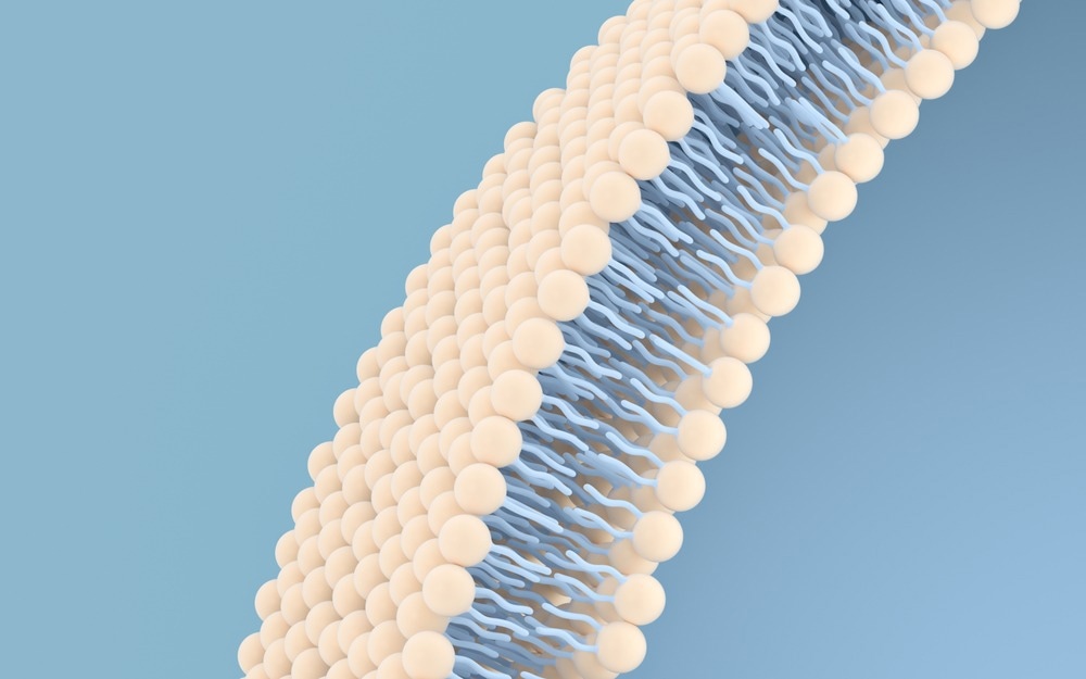
Study: Correct identification of the core-shell structure of cell membrane-coated polymeric nanoparticles. Image Credit: Vink Fan/Shutterstock.com
Recently, scientists analyzed several polymeric core nanoparticles using the fluorescence quenching method and demonstrated that some of the observed core-shell structures were actually artifacts created by the staining process. This study is available in Chemistry A European Journal.
The Role of Biomimetic Nanoparticles
Biomimetic nanoparticles containing functional CM coatings are effectively used in therapeutics as it offers desirable biological properties. Although several NPs, such as liposomes, iron oxide, gold, porous silica, and metal-organic frameworks (MOFs), were screened and treated with CM, only poly (lactic-co-glycolic acid) (PLGA) received approval from the United States Food and Drug Administration (FDA) for the production of CM-coated NPs.
Some of the characteristic features of PLGA are superior biodegradability, excellent drug loading capacity, and significant biocompatibility.
PLGA NPs (polymeric nanoparticle) have been applied in several therapeutics, such as red blood CM-coated anisotropic PLGA NPs used for detoxification, platelet membrane-coated PLGA NPs designed to improve cancer immunotherapy, and genetically engineered CM- camouflaged PLGA NPs for targeting inflammation.
Negative Staining and Development of Core-Shell Nanostructures
When CM materials and core NPs are exposed to ultrasonic energy or other disrupting forces, they are immediately transformed into an integrated core-shell nanostructure. This concept emerged from TEM images of CM-coated PLGA (CM-PLGA) NPs that are stained negatively.
Negative staining is associated with using an aqueous solution of electron scattering heavy-metal salts, such as phosphotungstic acid and uranyl acetate. These salts are deposited on the dried sample, which enhances its visualization. The negative straining method helps preserve the sample by forming a specimen mold. Nevertheless, some negative staining methods (e.g., air-drying negative staining) are inclined to develop artifacts that are often misinterpreted during TEM image analysis.
Accurate Detection of CM-PLGA NPs
Typically, techniques such as sodium dodecyl sulfate-polyacrylamide gel electrophoresis (SDS-PAGE), dynamic light scattering (DLS), and fluorescence co-localization are used to detect the presence of CMs on PLGA NPs. Scientists have recently sought to analyze if polymeric NPs are truly coated with CM, as previous TEM images with negative staining indicated.
In-depth research revealed that the core-shell nanostructure, visualized in TEM analysis of negatively stained polymeric NPs, which are commonly used as a core for CM coating, e.g., poly(caprolactone) (PCL), PLGA, and methyl ether-block-PLGA (PEG-PLGA), is an artifact created by the staining technique.
The co-fusion of CM and PLGA NPs was clarified using mouse colon carcinoma cells. Confocal laser scanning microscopic images confirmed the formation of CM-PLGA NPs. To determine the structure of CM-PLGA NPs, fluorescence quenching technique along with TEM analysis coalesced with Triton X-100 (TX-100) treatment was used.
Scientists compared TEM images of PLGA NPs with and without negative straining to accurately determine CM coating. Interestingly, the uranyl acetate stain revealed that most of the PLGA NPs possessed a core-shell structure. To ascertain the native state of PLGA NPs, which is commonly misinterpreted by the TEM imaging technique, cryogenic TEM (cryo-TEM) was used, as this method is popularly considered artifact-free.
Interestingly, the cryo-TEM images revealed a solid structure of PLGA NPs, which indicated that the assumed core-shell structure of the negatively stained PLGA NP’s TEM image could be an artifact. This artifact might be created during the staining and drying process. Atomic force microscopy (AFM) images of PLGA NPs exhibited smooth surface and spherical shape, which was consistent with field-emission scanning electron microscopy (FE-SEM) images.
Interestingly, the core-shell artifact was observed using both uranyl acetate and phosphotungstic acid negative stain, which suggested that core-shell artifacts were formed irrespective of the type of negative stain used. Alterations in the staining timing also did not impact the thickness of artifacts.
Conclusions
Taken together, most of the polymeric NPs, including PLGA, tend to falsely appear to have a core-shell structure in TEM analysis of negatively stained samples. This study confirmed that the core-shell structure of CM-PLGA NPs identified in several TEM images of negatively stained specimens are actually artifacts created by the staining of the original PLGA NPs and not due to CM coating.
Reference
Liu, L., et al. (2022) Correct identification of the core-shell structure of cell membrane-coated polymeric nanoparticles. Chemistry A European Journal. https://doi.org/10.1002/chem.202200947
Disclaimer: The views expressed here are those of the author expressed in their private capacity and do not necessarily represent the views of AZoM.com Limited T/A AZoNetwork the owner and operator of this website. This disclaimer forms part of the Terms and conditions of use of this website.