A common belief is that tumorigenesis and cancer progression represents a multistep process. The repeatedly used process for cancer diagnosis and prognosis to direct treatment decisions is formed on a multifaceted mixture of imaging and invasive tissue biopsies.
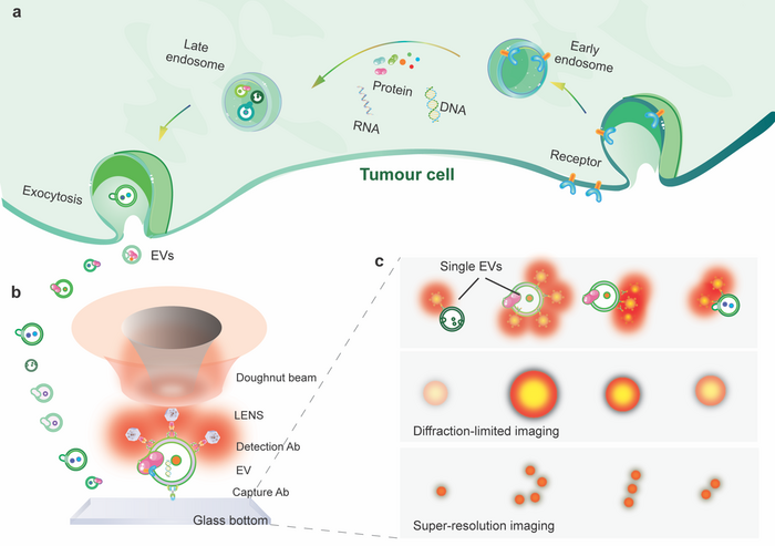
(a) sEVs carrying heterogeneous distribution of biomarkers are released from the tumor cell. (b) sEVs can be captured on an antibody-coated plate and labeled by UCNPs. (c) UCNPs -EV conjugates detected as single bright spots with different intensities can be super-resolved in super-resolution nanoscopy. Image Credit: Guan Huang, Yongtao Liu, Dejiang Wang, Ying Zhu, Shihui Wen, Juanfang Ruan, Dayong Jin.
The approaches are not always dedicated to early-stage cancer diagnosis. Small extracellular vesicles (sEVs) are nanometer-sized, bilayer lipid carriers and comprise a wide range of cargos, including proteins, lipids, metabolites, DNAs, and RNAs,
sEVs discharged from original cancer cells are found in nearly all body fluids. They can become the probable circulating biomarkers in liquid biopsies, as they exclusively reflect the dynamic biological variations connected with the emergent tumors and specify the stages of cancer progression.
Super-resolution microscopy methods have forced the resolution beyond the diffraction limit toward nanometer scales.
A team of researchers, guided by Professor Dayong Jin from the University of Technology Sydney, created an innovative technology established on Lanthanide-doped EV-targeting Nanoscopic Signal-amplifiers (LENS). Their article has huge potential in the diagnosis and prognosis of cancer and has been published in eLight.
Synthetic upconversion nanoparticles (UCNPs) have non-linear photo-switchable features. They allow a new type of super-resolution nanoscopy to realize an optical resolution of sub-30 nm.
The team’s latest work using nanophotonic probes further realized ultra-sensitivity in the quantitative uncovering of sEVs. These probes recorded approximately three orders of magnitude sensitivity better than the typical enzyme-linked immunosorbent assay (ELISA).
The scientists also enhanced the imaging resolution to super-resolve the surface biomarkers on single EVs. The method depends on using even, bright, and photostable nanophotonic probes.
Each is extremely doped with tens of thousands of lanthanide ions. During their experiment, the sEVs were first trapped on a slide coated with CD9 antibody and sandwiched by a biotinylated EpCAM antibody. Then, streptavidin functionalized upconversion nanoprobes tagged the EpCAM antibody for signal enhancement.
The nanoprobes on single sEVs facilitate a super-resolution microscope for visualization under a doughnut-shaped laser beam. A single nanoprobe at the center of the doughnut beam produces an emission pattern with a dip where the probe is positioned. The two adjacent nanoprobes can be super-resolved outside the diffraction boundary in the nanoscale.
The scientists show that super-resolution imaging of single sEVs can be accomplished using an array of upconversion nanoprobes doped with several kinds and diverse concentrations of emitters. They validate that antibody-conjugated nanoprobes can target tumor epitope epithelial cellular adhesion molecule (EpCAM) on large EVs and single sEVs.
Employing super-resolution imaging, the team could measure the exact number of nanoprobes on each sEV. They have demonstrated that it is hypothetically possible to examine nanoprobes’ size and steric interference on single sEVs.
Journal Reference
Huang, G., et al. (2022) Upconversion nanoparticles for super-resolution quantification of single small extracellular vesicles. eLight. doi.org/10.1186/s43593-022-00031-1.
Source: http://english.ciomp.cas.cn/