A new technique for imaging nanoparticles has been created by a team led by Professors Jinyang Liang and Fiorenzo Vetrone from the Énergie Matériaux Télécommunications Research Centre at the Institut national de la research scientifique (INRS). It is based on a high-precision, short-wave infrared imaging method that can record rare-earth doped nanoparticle photoluminescence lifetimes in the micro-to millisecond range.
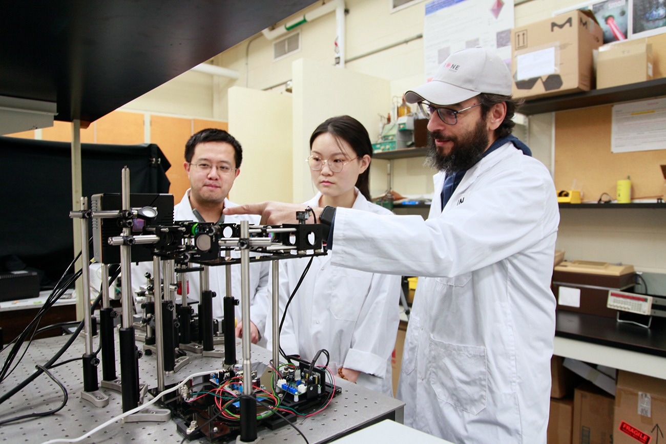 Jinyang Liang, INRS professor, Miao Liu, INRS PhD student in energy and materials science and Fiorenzo Vetrone, INRS Professor, in front of the SWIR-PLIMASC device. Image Credit: Institut National De La Recherche Scientifique
This innovative finding, which was detailed in the journal Advanced Science, opens up several exciting applications, especially in the information security and health domains.
Strategic metals, known as rare-earth elements, have special light-emitting qualities that make them highly desirable as research instruments in modern science. Furthermore, there is a benefit to the photoluminescence lifetime of these ion-doped nanoparticles being slightly impacted by external factors. Therefore, measuring it with imaging yields data that can be used to infer precise and extremely trustworthy information.
The optical technologies available for this kind of measurement were not optimal, notwithstanding the great advances being made in this field.
Until now, existing optical systems have offered limited possibilities due to inefficient photon detection, limited imaging speed, and low sensitivity.
Jinyang Liang, Professor, Institut National De La Recherche Scientifique
Jinyang Liang is also a specialist in ultrafast imaging and biophotonics,
Counting time-correlated single photons has been the most widely used method to date for determining the photoluminescence lifespan of rare-earth doped nanoparticles.
This method requires a large number of repeated excitations at the same location because the detector can only process a limited number of photons for each excitation.
Miao Liu Ph.D., Study First Author, Department of Energy and Materials Science, Institut National De La Recherche Scientifique
However, the repetition rate of the excitation is limited by the extended photoluminescence lifetimes of rare-earth doped nanoparticles in the infrared spectrum, which range from hundreds of microseconds to several milliseconds. Consequently, a significantly longer pixel staying time is required to construct the photoluminescence intensity decay curve.
Pushing the Limits
The teams led by Liang and Vetrone have used streak optics and a high-sensitivity camera to solve this obstacle. The end product is referred to as SWIR-PLIMASC (PLIMASC for photoluminescence lifetime imaging microscopy with an all-optical streak camera and SWIR for short-wave infrared).
Mapping the optical characteristics of short-wave infrared photoluminescence lifetimes is greatly enhanced by it. In the discipline of optics, it is the first high-speed, high-sensitivity SWIR imaging system.
Miao Liu said, “It has several advantages, for instance, it responds to a wide spectral range, from 900 nm to 1700 nm, allowing photoluminescence to be detected at different wavelengths and/or spectral bands.”
The Ph.D. student added that with a 1D imaging speed that can be tuned from 10.3 kHz to 138.9 kHz, photoluminescence lifetimes in the infrared spectrum, from microseconds to milliseconds, can be directly captured in one snapshot with the aid of this device.
Ultimately, assigning the photoluminescence's temporal information to various spatial coordinates guarantees that the whole 1D photoluminescence intensity decay process may be captured in a single photograph, devoid of repetitive excitation. Miao Liu said, “You save time, but still get high sensitivity.”
Biomedical and Security Applications
There will be a very noticeable result from the work done for this investigation. Professor Fiorenzo Vetrone, a nanomedicine specialist, thinks that SWIR-PLIMASC's advancements in the biomedical field could be used to combat cancer.
As our system applies to the temperature-based photoluminescence lifetime imaging of rare-earth ions, we believe that the data obtained could, for example, help to detect cancer cells even earlier and more accurately. The metabolism of those cells raises the temperature of the surrounding tissues.
Fiorenzo Vetrone, Professor, Institut National De La Recherche Scientifique
Fiorenzo Vetrone is also an expert in nanoparticles doped with rare earths.
The cutting-edge device may also be used to store data and documents at higher security levels precisely to stop them from being altered. Ultimately, these remarkable findings in basic science will enable researchers to create rare-earth nanoparticles with even more intriguing optical characteristics.
Journal Reference:
Miao Liu, et.al., (2024). Short-wave Infrared Photoluminescence Lifetime Mapping of Rare-Earth Doped Nanoparticles Using All-Optical Streak Imaging. Advanced Science. doi.org/10.1002/advs.202305284.
Source: https://inrs.ca/en/