In situ transmission electron microscopy (TEM) can be used to assess the chemical behavior of carbon nanotubes (CNTs), measuring nanoscopic characteristics and atomic-scale mechanisms.
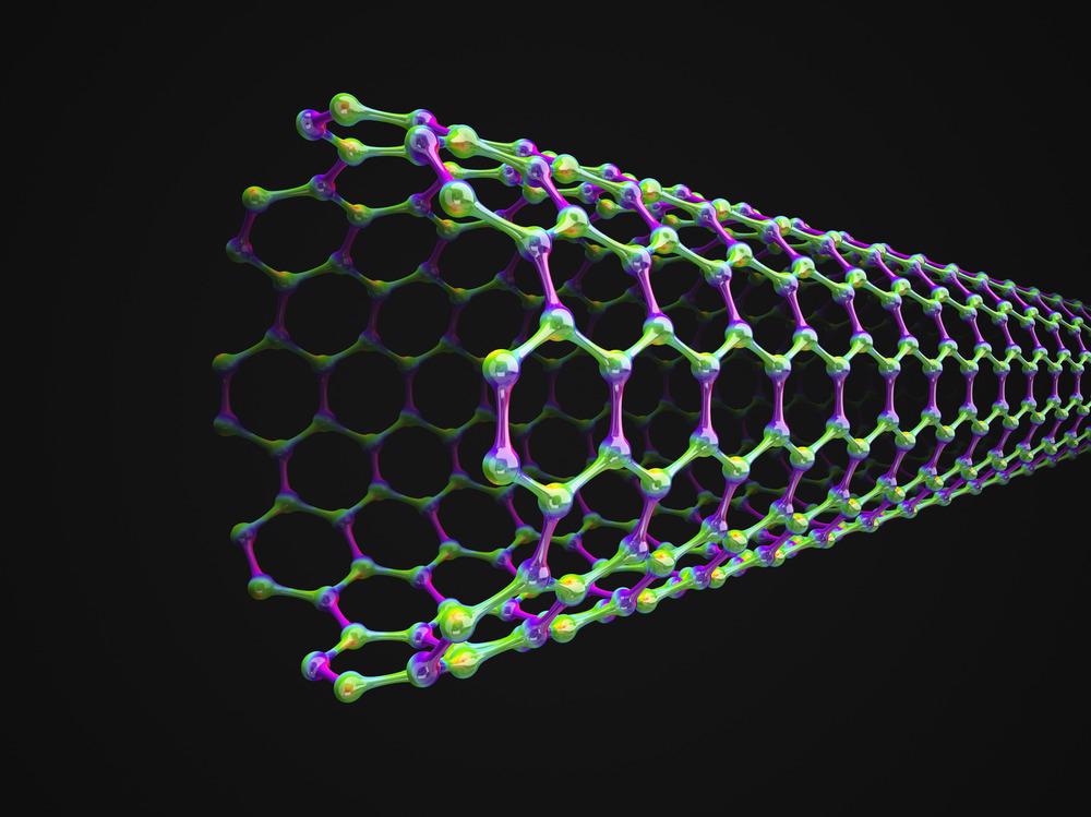
Image Credit: Orange Deer studio/Shutterstock.com
An Introduction to Carbon Nanotubes
Carbon nanotubes (CNTs) are 1D tube-like structures with nanometric diameters and micrometric length.
A carbon nanotube is made by the concentric rolling up of one or more graphene layers. These nanotubes may be categorized as multi-wall CNTs (MWCNTs), double-wall CNTs (DWCNTs), or single-wall CNTs (SWCNTs) based on the number of constituent graphene layers.
CNTs have been actively researched in various applications, ranging from fundamental scientific phenomena like quantum dot behavior to technology such as chemical sensors, nanoelectronics components, and electromechanical resonators.
Due to their distinct combination of properties like superior mechanical, electrical, and thermal qualities, CNTs have a lot of prospective applications in sensors, actuators, military equipment, robotic devices, and energy storage devices.
What is In Situ Transmission Electron Microscopy?
In situ transmission electron microscopy (TEM) is a technology that enables scientists to examine materials in real-time under real-life settings.
Since in situ methods overcome the vacuum-condition constraint of TEMs, scientists may retrieve far more information from specimen materials than they could with classical imaging techniques.
In Situ TEM Vs. Classical TEM
In situ TEM improves on the constraints of classical TEM. A transmission electron microscope is an observation-based instrument that is mainly meant to take a "picture" of a stationary specimen, but in situ approaches add testing possibilities to the microscope, making it a valuable approach for many researchers.
Scientists may manipulate the conditions in which their specimens are seen using micro-electro-mechanical system (MEMS) devices and in situ TEM holders, allowing them to execute actual experimentation inside the TEM. In the upcoming years, in situ TEM is expected to enable new research fields and be involved in many scientific breakthroughs.
The Advantages of In Situ TEM
While numerous specimens are invariably required for the imaging process in conventional TEM, a singular specimen may be utilized to perform tens of diverse in situ studies, increasing lab efficiency by producing more findings. In situ TEM may also be utilized for energy-dispersing X-ray spectroscopy (EDS) studies. This technique, which uses X-ray stimulation, enables comprehensive chemical evaluation of materials.
The use of solitary or mixed stimuli at the distinct nanostructure scale, in conjunction with concurrent microscopy and spectroscopy assessment, is a distinct benefit of TEM over conventional characterizing approaches.
In situ TEM has lately become much more accessible thanks to the employment of MEMS devices to regulate specimen settings on a Si chip of millimetric scale.
Such chips are inserted inside the TEM holder and are tailored to generate different real-world settings within the TEM, according to the researcher's objectives. In situ TEM holders, for instance, may generate heat, impart atmospheric pressure, and enable for liquid microscopic research.
Application of In Situ TEM in Carbon Nanotube Research
TEM uses have transitioned from post-mortem evaluation to in situ or live investigations of the morphology, chemistry, and characteristics of nanostructured materials. The development of innovative technologies, including rectification of aberrations and MEMS device incorporation, has significantly increased the pace of this paradigm shift.
From studying the catalytic nucleation and expansion of carbon nanotubes utilizing gas-solid interactions and elevated temperature radiation to mechanical characteristics evaluated via direct-force or indirect-force assessment methods, the employment of in situ TEM has aided in interpreting the reaction of CNTs to a variety of stimulants.
The iron-catalyzed MWCNT proliferation and cessation mechanisms were studied (Huang, Farra, Schlögl, & Willinger, 2019) using in situ TEM at nearly atmospheric pressures. The provided in situ TEM studies gave an atomic-level perspective of carbon nanotubes' proliferation and cessation processes under more realistic settings. It might prove to be a valuable guide for the controllable fabrication of CNTs by optimizing the catalytic design as well as the experimental conditions.
Future Prospects of In Situ TEM
In situ TEM investigations give a valuable understanding of real-life settings because of their adaptability and utility. With additional advancements in equipment, it is expected that the transmission electron microscope will soon emerge as one of the primary instruments for conducting chemical and physical research in individual nanomaterials like carbon nanotubes.
When combined with proper regulation and simulation studies, in situ TEM can give an atomically detailed understanding of reactions and physical responses, transforming it into a full-fledged nanoscale laboratory.
Equipment Upgrades Needed for Effective In Situ TEM
A basic device is required for effective in situ assessments: a TEM or a scanning transmission electron microscope (TEM/STEM) that integrates excellent spectral and spatial resolutions and may be linked with peripherals for in situ investigations.
Specimen holders able to apply extrinsic stimuli like strain, heat, cooling, electric bias, responsive settings (gas or liquid reaction cells), and photons are examples of peripherals. Furthermore, a data collecting and processing device able to enhance temporal resolution and manage massive data volumes would also be required.
The suggested equipment enhancements may generate huge volumes of imaging and spectroscopy data, necessitating fast data collection and transmission speeds.
References and Further Reading
Costa, P. M., & Ferreira, P. J. (2015). Advanced Transmission Electron Microscopy. In F. M. Deepak, Advanced Transmission Electron Microscopy (pp. 207-247). Springer. Available at: https://doi.org/10.1007/978-3-319-15177-9_7
Huang, X., Farra, R., Schlögl, R., & Willinger, M.-G. (2019). Growth and Termination Dynamics of Multiwalled Carbon Nanotubes at Near Ambient Pressure: An in Situ Transmission Electron Microscopy Study. Nano Letters, 19, 5380-5387. Available at: https://doi.org/10.1021/acs.nanolett.9b01888
Liu, C., & Cheng, H.-M. (2013). Carbon nanotubes: controlled growth and application. Materials Today, 16, 19-28. Available at: https://doi.org/10.1016/j.mattod.2013.01.019
Moering, J. (2017, November 22). What are the Advantages of In Situ TEM? Retrieved from Protochips: https://www.protochips.com/blog/advantages-of-in-situ-tem/
Taheri, M. L., Stach, E. A. et al (2016). Current status and future directions for in situ transmission electron microscopy. Ultramicroscopy, 170, 86-95. Available at: https://doi.org/10.1016/j.ultramic.2016.08.007
Disclaimer: The views expressed here are those of the author expressed in their private capacity and do not necessarily represent the views of AZoM.com Limited T/A AZoNetwork the owner and operator of this website. This disclaimer forms part of the Terms and conditions of use of this website.