Apr 1 2010
Since more than twenty years, scanning force microscopes are employed in research and industry. Their enormous resolution triggered many applications in nanotechnology. Their rather low image rate is however a disadvantage - changing objects and processes cannot be imaged.
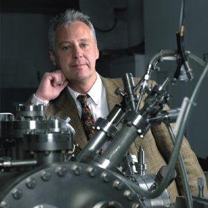 Physicists of Saarland University in the team of Prof. Uwe Hartmann (photo) have developed a technology that will accelerate scanning probe microscopes by a factor of 1000. photo: Bellhäuser - das bilderwerk
Physicists of Saarland University have developed a technology that could accelerate scanning probe microscopes by a factor of 1000. The operation principle is explained from April 17th to 23rd on the Saarland Forschungsstand on the Hannover Messe (Halle 2, Stand C 44).
A scanning probe microscope works like a record player. There, a needle follows the record track, mapping the fine structure of the track. The microscope uses a much smaller silicon needle instead, and direct contact with the surface is avoided. Surface structures are mapped by atomic forces, usually van-der-Waals interactions. "Even though the needle is tiny, there are still physical limits. Therefore we were looking for a component that is again a factor of 100 smaller than those used currently" explains Uwe Hartmann, Professor for Nanostructure Reasearch and Nanotechnology at Saarland University. With the nanocantilever, as it is called, surfaces will be mapped a lot faster and with higher precision.
State-of-the-art scanning probe microscopes operate at frequencies around 100 Kilohertz. "The processes nanotechnology is dealing with, however, have typical frequencies of gigahertz. These are one billion cycles per second. On the other hand, the velocity by which a hair is growing may well disturb the imaging process." Such are the dimensions of nanoresearch, as Uwe Hartmann describes. With his team's design, one hundred images per second and more and an increase in resolution will be possible. This is more than video rate.
The detector for the movements of the nanocantilever is separated from the nanocantilever by less than the wavelength of light, just one-fivehundredth of a hair's diameter. The result is a mapping of the surface with superior speed and precision.
In cooperation with partners a prototype of the new scanning force microscope is currently set up currently, for which is also patent application is intended. Until the end of the year the device, which uses only standard materials of microelectronics, will operate. The researchers are now looking for an industry partner. "On the Hannover Messe we will not show an exhibit. However we will demonstrate the principle of the scanning force microscope in a three-dimensional visualization" the researcher from Saarbrücken explains.