Researchers at the European Molecular Biology Laboratory (EMBL), Heidelberg, have developed a new microscope that helps to observe molecules’ movement in live cells for every millisecond.
 The new microscope enables scientists to watch and measure fast-moving molecules
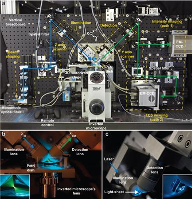
The new microscope integrates single molecule spectroscopy and light-sheet microscopy. This enables it to observe the fluorescence of each pixel within observation range and take pictures at time intervals below a millisecond. By this, the researchers can observe and measure extremely rapid processes including the pattern of diffusion of molecules across an entire sample even if it consists of numerous cells. This is a notable advancement as compared to past techniques using confocal microscopy where scientists could observe only some isolated spots in the sample simultaneously.
Until recently, chromatin which is a combination of chromosome-forming proteins, RNA and DNA were found in two states known as heterochromatin and euchromatin. Heterochromatin represents tightly packed DNAs, wherein most of the DNAs were not accessible to the gene-reading machinery of the cell. Euchromatin represents the loosely packed DNAs and allow easy readability. The researchers came up with an exciting finding when they measured the interaction between HP1-a, a protein, and chromatin using the newly developed microscope.
Michael Knop stated that in certain areas that seem as euchromatin, HP1-a acts in a manner which is similar to the way it would behave in the presence of heterochromatin. Hence it is now possible to observe chromatin in an intermediate state, that is between euchromatin and heterochromatin, which was impossible before in live cells. Malte Wachsmuth, developer of the microscope at EMBL stated that fluorescently-tagged molecules can be now be tracked in three-dimensional form in living cells and the variation of their biochemical properties such as binding affinities and interaction rates of the cell can be viewed with this microscope.
Scientists hope that this tool will favor in the analysis of processes that range from growth hormones role in tumors to cell division regulation as well as patterning and signaling of embryo’s tissue development.