Mar 10 2015
The major limitation of microscopy is light itself: if we want to see an object smaller than the wavelength of visible light, we cannot use conventional optics. When it comes to imaging cells, the problems compound, since cells often require chemical processing beforehand in order to make them suitable for viewing under a microscope.
This processing essentially kills the cell in order to preserve it. An EPFL spin-off company, NanoLive, has developed the 3D Cell Explorer first-ever microscope that allows users to see inside living cells without any prior sample preparation, by using MRI-like technology and proprietary software that uses holographic algorithms.
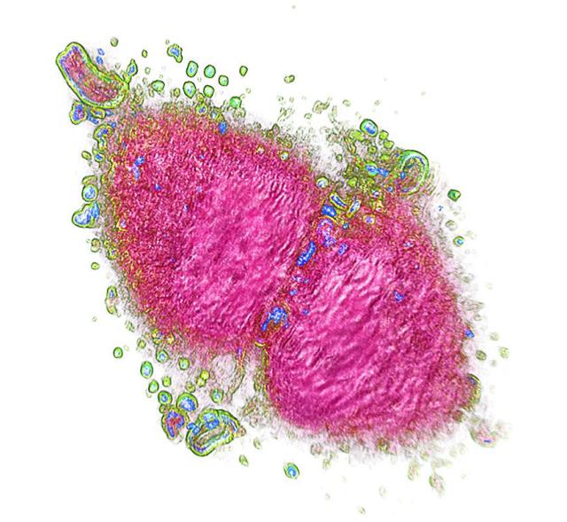 NanoLive, an EPFL spin-off, has brought to the market a new MRI-like microscope that can "see" into living cells without any previous processing. CREDIT: Nanolive
The microscope is called the 3D Cell Explorer. It combines state-of-the-art hardware with cutting-edge imaging software to record stunning 3D images of entire living cells within seconds and with a higher resolution than any conventional microscope available in the market. The device works as an MRI scanner, taking photographs at different depths across the cells. The photographic "slices" are then recombined using clever holography software that digitally "stains" the cells, labeling its different parts. The result is a high-resolution 3D image of the cell that can be rotated and explored in depth.
The software, called STEVE, is now available for download and can be used to "travel" virtually inside cells. It allows the user to digitally "paint" any part of a scanned cell. This is possible because it can automatically define all the different parts of a cell based on an optical property called the "refractive index". Since different organelles inside the cell have different refractive indices, STEVE can tell them apart and digitally "stain" them with the same or different colors. Because this digital processing is quantitative, it can use a limitless amount of colors, allowing users to explore changes in the cell in real time. Through STEVE, the stains can be constantly modified by the user, saved and reused for other cells.
"Starting from today, scientists, medics and students all around the world will be enabled to travel inside 3D cells in full color by simply downloading STEVE on their laptop" says NanoLive CEO Yann Cotte. Nanolive SA was founded in November 2013 at the EPFL Innovation Park (Lausanne) by Yann Cotte (CEO) and Fatih Toy (scientific advisor), who had just finished their PhDs at EPFL. Currently based in Ecublens, the company pioneered the 3D Cell Explorer in 2013, publishing their invention in Nature Photonics.
Source: http://www.epfl.ch/