Massachusetts Institute of Technology (MIT) engineers have designed a new atomic force microscope (AFM) that has the capability to scan images 2,000 times faster than models currently on the market. The microscope enables nearly real-time video of nanoscale chemical processes.
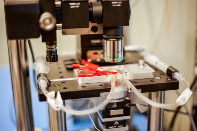 A new high-speed microscope produces images of chemical processes taking place at the nanoscale, at a rate that is close to real-time video. This closeup shot of the microscope shows transparent tubes used to inject various liquids into the imaging environment. This liquid can be water, acid, buffer solution for live bacteria, cells, or electrolytes in an electrochemical process. Researchers use one as an inlet and the other as an outlet to circulate and refresh the solutions throughout an experiment. (Photo: Jose-Luis Olivares/MIT)
Advanced AFMs have the ability to capture nanometer-scale images, and they have delivered close-up images of single DNA strands, hydrogen bonds and other atomic-sized structures. However, scanning such images is a long process and takes up a lot of time. AFMs have been predominantly used for imaging static samples as they are not able to capture changing environments.
Using the newly developed AFM, the team scanned a 70 x 70 µm calcite sample as it was immersed in deionized water and exposed to sulfuric acid, and they observed the processes involved – the calcite being eaten by acid, the nanometer-sized pits expanding and rapidly merging, and the removal of calcite layer-by-layer along the crystal pattern of the material within seconds.
Kamal Youcef-Toumi, a professor of mechanical engineering at MIT, stated that the sensitivity and speed of the AFM could help observe atomic-sized processes taking place in a high-resolution “movie” format.
Microscope creates near-real-time videos of nanoscale processes
“People can see, for example, condensation, nucleation, dissolution, or deposition of material, and how these happen in real-time — things that people have never seen before,” Youcef-Toumi says. “This is fantastic to see these details emerging. And it will open great opportunities to explore all of this world that is at the nanoscale.”
The design and images that are part of the study are based on postdoc Iman Soltani Bozchalooi’s PhD work.
In AFMs, an ultrafine needle or probe traces the topography of a sample by skimming along the sample’s surface. Samples are placed on a movable platform that moves beneath the probe laterally and vertically. As the structures are extremely small, and the instruments have to function very slowly, avoiding abrupt movements to prevent the image from being blurred or the sample from being modified. The typical speed of these microscopes is approximately one to two lines per second.
“If the sample is static, it’s ok to take eight to 10 minutes to get a picture,” Youcef-Toumi says. “But if it’s something that’s changing, then imagine if you start scanning from the top very slowly. By the time you get to the bottom, the sample has changed, and so the information in the image is not correct, since it has been stretched over time.”
Researchers have attempted to increase the speed of the scanning process by building smaller, nimbler, and more rapidly scanning platforms, but such scanners do not allow a wider view or zoom out.
“It’s like if you were landing somewhere in the United States and have no clue where you’re landing, and are told wherever you land, you’re only allowed to look a few blocks around and up to a limited height,” Bozchalooi says. “There’s no way you can get a bigger picture.”
Bozchalooi developed a design that enables rapid scanning over smaller and larger ranges. The most important part is a multiactuated scanner and its control. The sample platform has both a larger, slower scanner and a smaller, speedier scanner for every direction. These function as a single system and enable scanning of wide 3D regions at high speed.
The interactions between scanners have been a significant factor that has thwarted development of multiactuated scanners. One scanner’s movement affects the motion and accuracy of the other. Furthermore, controlling each scanner separately and enabling them to work with all other microscope components has proven to be difficult. To scan every sample, multiple tunings and adjustments have to be made to numerous components in the AFM instrument.
Bozchalooi has developed control algorithms that address these problems and enable the multiactuated instrument to be used easily.
“Our controller can move the little scanner in a way that it doesn’t excite the big scanner, because we know what sort of motion triggers this scanner, and vice versa,” Bozchalooi says. “In the end, they’re working in synchrony, so from the perspective of the scientist, this scanner looks like a single, high-speed, large-range scanner that does not add any complexity to the operation of the instrument.”
The team optimized the optics, instrumentation, data acquisition systems and other components of the AFM. They observed that the instrument had the capability to scan a calcite sample both forward and backward, without the sample or AFM probe getting damaged. When compared to existing commercial AFMs, the new AFM scans 2,000 times faster, at a rate of more than 4,000 lines per second, 2,000 Hz, or approximately 8 to 10 frames per second.
Bozchalooi states that the instrument would not have any imaging range limit, and it would be able to scan features with a height of several microns, and across hundreds of microns.
“We want to go to real video, which is at least 30 frames per second,” Youcef-Toumi says. “Hopefully we can work on improving the instrument and controls so that we can do video-rate imaging while maintaining its large range and keeping it user-friendly. That would be something great to see.”
The study has been published in the journal, Ultramicroscopy.
The Center for Clean Water and Clean Energy at MIT and KFUPM, and National Instruments supported the research.