Dec 10 2019
For many years, it had remained an inexplicit dream to analyze the single-particle dynamics at the femtosecond level and nanoscale.
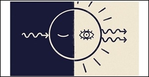 A single photon stimulates emission of a second, twin photon from a quantum dot, making the quantum dot detectable and revealing its excited state dynamics. Image Credit: Joanna Ambroz.
It was only at the beginning of the 21st century that femtoscience and nanotechnology were slowly combined together, and the initial ultrafast microscopy of individual molecules and quantum dots (QDs) was eventually achieved.
Studies relating to ultrafast microscopy fully depend on identifying single molecules or nanoparticles using luminescence methods, which work only through efficient emitters. But these methods degrade the sample and also yield only minimal information regarding the dynamics of the system in the activated state.
Attempts to identify another compatible method to analyze rapid processes in nano-objects were discovered only in the recent past.
At ICFO, a research team, including Lukasz Piatkowski, Nicolò Accanto, Gaëtan Calbris, and Sotirios Christodoulou, in association with Iwan Moreels (Ghent University in Belgium), has now developed a new method for analyzing ultrafast events in each non-fluorescent nano-object.
The study titled “Ultrafast stimulated emission microscopy of single nanocrystals” has been published in the Science journal. It was headed by ICREA Professor Niek F. van Hulst at ICFO.
The researchers considered individual QDs in their latest study. But instead of waiting for the individual QDs to suddenly discharge light via photoluminescence, they encouraged the individual QDs into an excited state using an advanced combination of laser pulses. These QDs were then forced down and brought back to the ground state to initially image individual QDs and subsequently distinguish the emergence of the excited charges inside the whole photocycle.
Dr Lukasz Piatkowski explained why a laser pulse pair was utilized to successfully image the QDs’ dynamics.
It is like throwing a ball into a tree; the higher you throw it, the more excited the state. The first laser pulse of the system (photon) throws the first ball (charge in the QD) into the tree. If you are using a photoluminescence technique it is like you are standing below the tree, and you cannot see what is happening inside the treetop or crown.
Dr Lukasz Piatkowski, Researcher, ICFO
Piatkowski continued, “Thus, you will not know whether the ball starts to bounce down the branches, where, when and how it starts to fall down, if it stomps with something on its way, if it gets caught in an intermediate branch, etc. So, in order to see what is happening with the first ball, you need to find another technique that allows you to look into the treetop.”
The technique we used allowed us to throw a second ball into the tree top (second laser pulse interacting with the QD) to bring the first ball down. Throwing the second ball higher or lower, stronger or weaker, sooner or later after the first ball, we obtain information about the first ball and the structure of the tree (how long it took the balls to fall out, where, how, etc.).
Dr Lukasz Piatkowski, Researcher, ICFO
In the researchers’ experiment, individual QDs were brought to the excited state by the first laser pulse. A second laser pulse was then shot onto the QDs every few hundred femtoseconds to bring the excited charges down to the ground state. This induces recombination and emission of an additional photon.
Therefore, when the researchers shot each probe photon into the system, they obtained a pair of twin photons back. These additional photons enabled the researchers to image the individual QDs and also to accurately monitor the emergence of the excited charges in the QDs. This revealed the number of charges that experienced stimulated recombination, spontaneous recombination, and excited-state absorption.
Nanoscale monitoring of excited charges is very important in photovoltaics, photonics, and nanotechnology. The study results have demonstrated that ultrafast processes in individual chromophoric particles can be studied using ultrafast stimulated emission microscopy. Such particles cannot be detected using photoluminescence or fluorescence methods.
To put this in simpler terms, an analysis like that has made it possible to image and investigate the dynamics of nanoparticles as well as structures without using any external fluorescent labels.
Significant advances are expected in the future within the field of ultra-fast-nano-regime imaging techniques. The first detection of quantum dots using this approach has been outstanding. We now aim to extend this to molecules and biomolecular complexes, specifically photo-synthetic complexes.
Niek van Hulst, ICREA Professor, ICFO
“We are currently working on 3 and 4 pulse schemes to merge the stimulated emission and luminescence detection of single systems with 2D-spectroscopy,” concluded van Hulst.
Source: https://www.icfo.eu/