Feb 25 2020
At the Paul Scherrer Institute (PSI), scientists have successfully captured a short “3D film” of magnetic processes on the nanometer level for the first time.
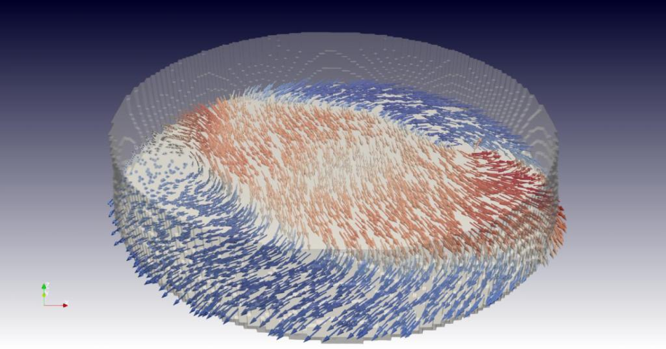 A look inside the material: a snapshot from the 3D film of the researchers. It shows a cross-section through the sample and in this plane the local alignment of the magnetic moments, represented by arrows in red (pointing to the right) and blue (pointing to the left). This makes the wavy structure and vortices in the magnetization visible. Image Credit: Paul Scherrer Institute/Claire Donnelly.
The breakthrough shows a wide range of dynamics within the material, such as the movement of swirling boundaries that exist between different magnetic domains. This new understanding was achieved with a recently developed technique at the Swiss Light Source (SLS). This approach could help make magnetic data storage devices more efficient and compact.
The scientists have recently published their study results in the Nature Nanotechnology journal.
It is hardly surprising to see a magnetic sticker that remains fixed to a refrigerator door. But when it comes to the nanometer scale (where 1 nm corresponds to one-millionth of a millimeter), magnets and their behavior are rather puzzling to physicists.
Meanwhile, the impacts that occur on this tiny scale are highly applicable to upcoming technologies. Now, PSI scientists have recorded a brief “film” of the three-dimensional (3D) magnetic structure within a material with nanoscale resolution, for the first time.
Magnetism plays a role in many ways in our everyday lives; but at this very small, fundamental level, the phenomena are not yet fully understood.
Claire Donnelly, Study Lead Author, University of Cambridge
Donnelly was a researcher at PSI when the experiment was performed and currently works at the University of Cambridge in the United Kingdom.
The scientists utilized X-ray light from the SLS at PSI and also employed a unique tomographic technique that they developed recently at the institute and dubbed “time-resolved ptychographic laminography.”
The researchers’ team included scientists at ETH Zurich, PSI, and also from the United Kingdom. The sample assessed by the team contained a gadolinium-cobalt compound that was patterned into a spherical disk.
More than Four Days for Seven Images
With our method we can non-destructively scan the material and from the data reconstruct several successive 3D images of the inner magnetic structure. We can visualise the orientation of the magnetic moment at every measured point in the material and represent them as tiny magnetic compass needles.
Manuel Guizar-Sicairos, Researcher, Paul Scherrer Institute
These compass needles respond to an external magnetic field and to one other, similar to the reaction of magnetic filings, and create complex patterns across the whole object. These patterns include the so-called domains, or areas, where the magnetization mostly points in one direction.
The changes that take place between two such domains—that is, the domain walls—hold specific interest for scientists: “People have proposed using them as memory bits, which could possibly be used to pack data even more tightly than when using the domains,” added Donnelly.
With the help of advanced imaging techniques, the specifics of these domain walls were only recently viewed in 3D at PSI, as well as at other locations. In the current analysis, the scientists moved one step ahead by mapping the movement of the domains as well as the domain boundaries.
“We have taken seven snapshots showing points in time that are only a quarter of a billionth of a second apart. In these we can see how a domain boundary moves back and forth,” added Donnelly. The researchers took slightly more than four days to continuously measure to gather the data, which subsequently produced this series of seven images.
Like Stroboscopic Light
With the help of an externally applied magnetic field, the scientists themselves induced the visualized motion of the domain boundary specifically and repeatedly. Hence, the researchers’ images were not exactly captured within a quarter of a billionth of a second.
The team instead produced a time loop of the varying magnetic field and captured images at different points in time within it, just like stroboscopic light that appears to slow down a repetitive motion.
Consequently, the recording of the 3D images from within the sample draws on a fundamental principle from computed tomography (CT). Just like medical CT scans, the X-rays were utilized to take several radioscopic images of the sample consecutively, each from a somewhat different angle.
From the data obtained, the scientists successfully recovered their 3D maps of the magnetization utilizing the software they had created just for this purpose.
With this method, we have not just achieved time-resolved 3D movies of the interior of an object. We also have been able to map the nanoscale dynamics in a magnet. In other words, we have shown that our new technique is really relevant to the development of new technology.
Claire Donnelly, Study Lead Author, University of Cambridge
“Our new method is also suitable for other materials and could therefore have many more useful applications in the future,” concluded Guizar-Sicairos.
Source: https://www.psi.ch/