Electron microscopy and analysis instruments from Carl Zeiss are redefining the science of brain mapping and 3-D reconstruction. Wide-field, high resolution imaging, huge data storage and manipulation capabilities plus highly automated sample preparation techniques are yielding research results 100-times, or more, faster than ever before. As a result, a number of high-ranking research institutes are adopting these remarkable instruments, and are producing phenomenal breakthroughs in understanding the human brain.
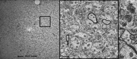 Specific region of the dentate gyrus of the hippocampus of a rat. 49µm x 49µm field of view, resolution two Nanometer.
For example, a University of Texas lab specializes in three-dimensional reconstruction by stacking images from serial 45nm-thick sections. According to John Mendenhall, a microscopist at the Center for Learning and Memory, their approach to accelerating the analysis is to “map the largest single field of view at minimum resolution, 2-3 nm pixels at the specimen level, needed for benchmarking ultrastructural features. With enough sections, sufficient resolution and the large field-of-view, we can approach neural circuit-scale volumes that include essential sub-cellular and connectivity detail.” Mendenhall said that the SUPRA™ FE scanning electron microscope from Carl Zeiss was chosen for this work because of its extreme field-of-view imaging. “The key is the super sampling frame store of images up to 32 thousand by 32 thousand pixels. This field-of-view can be a more than a hundred times larger area than with a standard frame store, which means that image acquisition and registration are much faster, and the resolution is retained below 4 nm.” In the meantime, even 64X64 k is supported by the system architecture of Carl Zeiss instruments, showing ongoing improvements in that area.
In addition, at the École Polytechnique Fédérale de Lausanne (EPFL), Dr. Marco Cantoni and his collaborators rely on a Carl Zeiss NVision 40 CrossBeam workstation for brain research. “We have produced thousands of slices from a mouse brain using the combination of high resolution SEM imaging and FIB slicing,” said Cantoni. “The results are incredible. Within a 48 hour nonstop machine run we generated a stack of 1600 images with 5 nm pixel size in X and Y direction and a FIB slice thickness of 6 nm. In virtual slices of the reconstructed volume we could not see any difference in resolution in the third dimension. The actual voxel size is about 5x5x6 nm—giving us an unprecedented insight into the three-dimensional structure of the investigated tissue.”
A wide variety of imaging and analysis instruments from Carl Zeiss are continuing to enable these accelerating results in the field of brain mapping.
Brain mapping
Attempting to understand the relationship between structure and function of the human brain is, perhaps, the hottest topic within the life science R&D community today. The results are critical. Lessons learned are immediately applicable to the design of surgical and medical interventions, and to the treatment of psychological and psychiatric disorders. The challenge is huge. With a hundred billion neurons responsible for a multitude of brain functions, figuring out how these cells interact seems an overwhelming task. But today’s new technology is speeding the process.
Posted on October 8th, 2009