Cryogenic-electron microscopy (cryo-EM) has rapidly become one of the most important experimental methods in structural biology and nanoscience. In this article, AZoNano discusses the working principle of single particle cryo-electron microscopy.
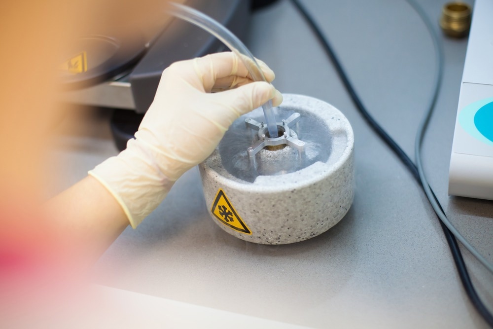
Image Credit: PolakPhoto/Shutterstock.com
The ability of single particle cryo-electron microscopy to capture structural information on samples that could not be crystallized for use with crystallographic methods or were too delicate for electron microscopy techniques has made it possible to look at a wealth of new structures and sample types.1,2
In this guide, we look at how single particle cryo-electron microscopy has evolved as a technique and, in particular, how it has been applied to look at problems in nanoscience.
Single Particle Cryo-Electron Microscopy: A History
Since the development of the first electron microscopes in the 1930s, electron microscopy methods have been incredibly important tools in the sciences. One of the key advantages of electron microscopy methods over older optical microscopy methods is the improved spatial resolution. For standard visible light microscopes, the diffraction limit of optical light is on the order of a few hundred nanometers, nearly twice the length of a typical carbon-carbon bond.
In contrast to this, electron microscopes can achieve sub-Angstrom spatial resolutions, making them suitable for resolving even the finest structural details in new nanomaterials.3
Cryo-EM involves the cryogenic cooling of samples to rapidly ‘freeze out’ their structure at a given moment in time. While the idea of freezing and slicing samples for microscopy has been around since the 1950s, one of the biggest challenges was the formation of crystalline ice in the freezing procedure.4 The ice crystals cause a large amount of electron diffraction, obscuring the key features in the image.
Improvements in sample freezing techniques to reduce the formation of water crystals, direct-beam detector sensitivities and 3D image reconstruction algorithms were some of the key developments that contributed to making it possible to record the first high-resolution 3D images of structures with single particle cryo-electron microscopy of samples that were not highly ordered.4
The significance of this development in single particle cryo-electron microscopy and its impact on many different scientific areas was ultimately recognized with the 2017 Nobel Prize in Chemistry.
Single Particle Cryo-Electron Microscopy: How It Works
Performing single particle cryo-electron microscopy measurements on nanoobjects has many similarities to other electron microscopy techniques, with the notable difference being the cryogenic sample preparation. Once the nanomaterial has been frozen into a glass-like state, it can be loaded into a transmission electron microscope.
The single particle electron microscopy instrument uses an intense beam of electrons focused through the microscope column using a series of electrostatic lenses and pass through the sample. The transmitted beam is then imaged onto a camera beneath the sample.
One important development for single particle cryo-electron microscopy was the ability to reconstruct full 3D models from recording a series of 2D images. This is done via image construction algorithms that combine 2D images of the sample in different orientations.
Some key considerations in single particle cryo-electron microscopy are avoiding sample damage with the intense electron beam and ensuring there is sufficiently good contrast in the final image.5 Improvements in camera technologies that meant weaker electron signals could be detected were critical in improving the scope of samples that cryo-EM was suitable for imaging.
The 2017 Nobel Prize in Chemistry: Cryo-electron microscopy explained
Video Credit: Chemical & Engineering News/YouTube.com
Single Particle Cryo-Electron Microscopy: Applications in Nanoscience
The key to the success of single particle cryo-electron microscopy in nanoscience is single particle cryo-electron microscopy's high spatial resolution. Single particle cryo-electron microscopy also has the advantage over other electron microscopy techniques of improved sample durability from cryogenically cooled species.6
Batteries have been one aspect of nanoscience that is being increasingly studied with single particle cryo-electron microscopy as many of the processes involved in the loss of battery efficiency are associated with nanoscale processes, such as the deactivation of lithium.7 Lithium has been historically very difficult to measure with electron microscopy methods. However, being able to spatially resolve processes such as dendrite formation on the nanoscale has made it possible to understand many previously unseen degradation processes.
Nanoparticle sizing is another application of single particle cryo-eletron microscopy as well as the profiling of biological and inorganic nanoscale structures.8 The life sciences have probably been the largest application area of single particle cryo-electron microscopy. Still, the development of low-dose single particle cryo-electron microscopy techniques means that more applications in materials science are now becoming feasible.6
The high spatial resolution of single particle cryo-electron microscopy and the ability to use image analysis methods to reconstruct higher dimensional images has also led to single particle cryo-electron microscopy’s use as a tool for imaging nanoreactors.
Nanoreactors are nanoscale objects, often inspired by biological proteins and enzymes, that can enhance specific chemical reactions for synthesis or catalysis. Often, nanoreactors are made by exploiting the size matching between specific chemical reactants or tuning the intermolecular interactions to bind particular substrates.
The success of nanoscale engineering, such as developing nanoplatforms to use as nanoreactors relies on characterization methods such as single particle cryo-electron microscopy that can characterize the spatial dimensions and structures of these nanoscale objects.
References and Further Reading
Callaway, E. (2020). The protein-imaging technique taking over structural biology. Nature, 578, p.201. https://media.nature.com/original/magazine-assets/d41586-020-00341-9/d41586-020-00341-9.pdf
Ju, Z., Yuan, H., Sheng, O., Liu, T., Nai, J., Wang, Y., Liu, Y., & Tao, X. (2021). Cro-Electron Microscopy for Unveiling the Sensitive Battery Materials. Small Science, 1, 210055.
Smith, D. J. (2008). Ultimate resolution in the electron microscope? Materials Today, 11, pp.30–38. doi.org/10.1016/S1369-7021(09)70005-7
Brzezinski, P. (2017). The Development of Cryo-Electron Microscopy. Available at: https://www.nobelprize.org/uploads/2018/06/advanced-chemistryprize2017.pdf
Baker, L. A., & Rubinstein, J. L. (2010). Radiation damage in electron cryomicroscopy. In: Methods in Enzymology (Vol. 481, Issue C). Elsevier Masson SAS. doi.org/10.1016/S0076-6879(10)81015-8
Li, Y., Huang, W., Li, Y., Chiu, W., & Cui, Y. (2020). Opportunities for Cryogenic Electron Microscopy in Materials Science and Nanoscience. ACS Nano, 14(8), pp.9263–9276. doi.org/10.1021/acsnano.0c05020
Fang, C., Li, J., Zhang, M., Zhang, Y., Yang, F., Lee, J. Z., Lee, M. H., Alvarado, J., Schroeder, M. A., Yang, Y., Lu, B., Williams, N., Ceja, M., Yang, L., Cai, M., Gu, J., Xu, K., Wang, X., & Meng, Y. S. (2019). Quantifying inactive lithium in lithium metal batteries. Nature, 572(7770), pp.511–515. doi.org/10.1038/s41586-019-1481-z
Živanović, V., Kochovski, Z., Arenz, C., Lu, Y., & Kneipp, J. (2018). SERS and Cryo-EM Directly Reveal Different Liposome Structures during Interaction with Gold Nanoparticles. Journal of Physical Chemistry Letters, 9(23), pp.6767–6772. doi.org/10.1021/acs.jpclett.8b03191
Hand, E. (2020). ‘We need a people’s cryo-EM’. Available at: https://www.science.org/content/article/we-need-people-s-cryo-em-scientists-hope-bring-revolutionary-microscope-masses
Peplow, M (2020). Cheaper cryo-EM on the horizon. Available at: https://cen.acs.org/analytical-chemistry/microscopy/Cheaper-cryo-EM-horizon/98/web/2020/11
Skolnick, J., Gao, M., Zhou, H., & Singh, S. (2021). AlphaFold 2: Why It Works and Its Implications for Understanding the Relationships of Protein Sequence, Structure, and Function. Journal of Chemical Information and Modeling, 61(10), pp.4827–4831. doi.org/10.1021/acs.jcim.1c01114
Disclaimer: The views expressed here are those of the author expressed in their private capacity and do not necessarily represent the views of AZoM.com Limited T/A AZoNetwork the owner and operator of this website. This disclaimer forms part of the Terms and conditions of use of this website.