Researchers at the Lawrence Berkeley National Laboratory (Berkeley Lab) and the University of California (UC) Berkeley, have discovered a new cathodoluminescence activated imaging by resonant energy transfer (CLAIRE) technique that extends high electron microscopy resolution to the dynamic imaging of soft matter.
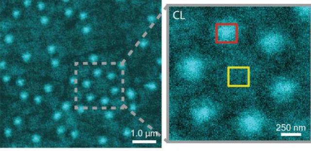 This is a CLAIRE image of Al nanostructures with an inset that shows a cluster of six Al nanostructures. Credit: courtesy of Naomi Ginsberg, Berkeley Lab
Soft matter covers a broad number of materials which includes polymers, liquids, foam, gels and the most significant is biomolecules. Nano-sized component interaction is at the core of soft materials and governs their capabilities and properties.
To gain an insight into important biological processes as protein metabolism and crystallization, it is important to observe the dynamics behind these interactions and this could help in improving the development of new technologies such as high-efficiency photovoltaic cells or artificial photosynthesis. It is a key challenge to observe these dynamics at considerable resolution and this has been made possible with a novel nanoscale imaging technique CLAIRE.
"Traditional electron microscopy damages soft materials and has therefore mainly been used to provide topographical or compositional information about robust inorganic solids or fixed sections of biological specimens," says chemist Naomi Ginsberg, who leads CLAIRE's development.
"CLAIRE allows us to convert electron microscopy into a new non-invasive imaging modality for studying soft materials and providing spectrally specific information about them on the nanoscale."
Ginsberg holds appointments with Berkeley Lab's Physical Biosciences Division and its Materials Sciences Division, as well as UC Berkeley's departments of chemistry and physics. She is also a member of the Kavli Energy NanoScience Institute (Kavli-ENSI) at Berkeley. Her team showed the imaging capabilities of CLAIRE by using the technique for aluminum nanostructures and polymer structures that could not have been imaged directly with electron microscopy.
"What microscopic defects in molecular solids give rise to their functional optical and electronic properties? By what potentially controllable process do such solids form from their individual microscopic components, initially in the solution phase? The answers require observing the dynamics of electronic excitations or of molecules themselves as they explore spatially heterogeneous landscapes in condensed phase systems," Ginsberg says.
"In our demonstration, we obtained optical images of aluminum nanostructures with 46 nanometer resolution, then validated the non-invasiveness of CLAIRE by imaging a conjugated polymer film. The high resolution, speed and non-invasiveness we demonstrated with CLAIRE positions us to transform our current understanding of key biomolecular interactions."
CLAIRE integrates into a single platform the best of scanning and optical electron microscopy. Scanning electron microscopes use electron beams instead of light for magnification and illumination. Electron beams can be used that have much shorter wavelengths when compared to photons of visible light for observing objects hundreds of times smaller than those that can be resolved using an optical microscope. These electron beams however destroy most soft matter forms and are not capable of spectrally specific molecular excitation.
Ginsberg and her team overcame this problem using "cathodoluminescence," in which a 20nm thick ultrathin scintillating film, made of cerium-doped yttrium aluminum perovskite, is sandwiched between the sample and the electron beam. When a low-energy electron beam of about 1keV excites a scintillating film, the energy emitted is transferred to the sample, causing the sample to radiate. The luminescence is measured and correlated to the electron beam position to form an image not limited by the optical diffraction limit.
Ginberg states that the development of the scintillating film and combining it into a microchip imaging device was a very important task and she applauds her research team for the success. She also credited the staff and the capabilities of a DOE Office of Science User Facility, the Molecular Foundry, where this CLAIRE imaging demonstration was performed.
"The Molecular Foundry truly enabled CLAIRE imaging to come to life," she says. "We collaborated with staff scientists there to design and install a high efficiency light collection apparatus in one of the Foundry's scanning electron microscopes and their advice and input were fantastic. That we can work with Foundry scientists to modify the instrumentation and enhance its capabilities not only for our own experiments but also for other users is unique."
Ginsberg and her team are working towards making CLAIRE widely accessible and customizing it further for particular applications.
"We're interested in non-invasively imaging soft functional materials like the active layers in solar cells and light-emitting devices," she says. "It is especially true in organics and organic/inorganic hybrids that the morphology of these materials is complex and requires nanoscale resolution to correlate morphological features to functions."
Ginsberg and her team are also working on creating liquid cells for the observation of biomolecular interactions under physiological conditions. As it is possible for electron microscopes to function only in a high vacuum, as the electron beam is disrupted by molecules in the air and as evaporation of liquids takes place in high vacuum, aqueous samples must either be hermetically sealed or freeze-dried in special cells.
"We need liquid cells for CLAIRE to study the dynamic organization of light-harvesting proteins in photosynthetic membranes," Ginsberg says. "We should also be able to perform other studies in membrane biophysics to see how molecules diffuse in complex environments, and we'd like to be able to study molecular recognition at the single molecule level."
Furthermore, the dynamics of nanoscale systems for soft materials in general will be studied using CLAIRE by Ginsberg and her team.
"We would love to be able to observe crystallization processes or to watch a material made of nanoscale components anneal or undergo a phase transition," she says. "We would also love to be able to watch the electric double layer at a charged surface as it evolves, as this phenomenon is crucial to battery science."
A paper titled "Cathodoluminescence-Activated Nanoimaging: Noninvasive Near- Field Optical Microscopy in an Electron Microscope" has been published in the Nano Letters journal. The corresponding author of the paper is Ginsberg. Other authors are Connor Bischak, Craig Hetherington, Zhe Wang, Jake Precht, David Kaz and Darrell Schlom.