Nanomedicine has demonstrated great efficacy in delivering an array of therapeutic compounds into the body's target sites. The use of nanomedicine in healthcare necessitates a thorough understanding of its morphological features, bio-distribution, localization, and stability. Here, we discuss the application of TEM in nanomedicine for drug discovery and targeting.
Image Credit: Billion Photos/Shutterstock.com
Characterization Techniques in Nano-Drug Delivery
Nano-drug delivery techniques have expanded the possibilities for delivering a broad spectrum of biomolecular and synthetic therapeutic agents into a body target region.
The characterization of various nanoformulations, particularly the examination of nanostructure, stabilization, and biological transit through the body, remains a key factor for the effectiveness of any drug delivery strategy. This necessitates the use of advanced characterization methods.
Chromatography, X-ray scattering, electrophoresis, spectroscopy, zeta potential testing, mass spectrometry, and microscopy are some of the analytical procedures used to evaluate physicochemical features.
TEM in Nanomedicine
Microscopy has considerable potential for detecting the underlying structures of nanoparticles (NPs), which govern their performance and uses. Microscopy-based approaches have been utilized to characterize NPs, as well as their proficient design and adoption of safe procedures to be used in nanomedicine.
Transmission electron microscopy (TEM) is a valuable and powerful tool for characterizing NPs. Its great resolution for structural features is crucial to acquiring information about the granularity and crystal structure of NPs.
The TEM technique is employed for characterizing and assessing NPs in the fabrication of nano-drug delivery systems. It is utilized to analyze potential changes in the shape of nanoparticles after drug integration at various doses.
What is the Principle Behind TEM?
TEM is equipped with an electric gun that emits thermoionic radiation using lanthanum hexaboride (LaB6) or tungsten. It sends an electron across a thin slice of specimen to generate a two dimensional (2D) representation on a phosphorescent screen. Electrons transmitted via the sample are proportional to its brightness.
The traditional TEM scans with a range of 60-100 keV, making it better suited for biological specimens. However, high-voltage electron microscopes (HVEMs) and high-resolution transmission electron microscopes (HRTEMs) operate at roughly 200 keV-3 MeV.
Applications in Drug Delivery
TEM is used to determine the shape, size, and morphology of nanoparticles. The TEM imaging has a significantly greater resolution and generates high-quality images.
A TEM micrograph can reveal the amount of nanoparticle exfoliation, intercalation, and orientation. TEM photographs can also be used to demonstrate the pattern of distribution and dispersal of NPs in applications like polymer matrices utilized in nanocomposite synthesis.
It can be used to analyze alterations to structure caused by their interactions with gas-, solid-, or liquid-phase substrates. It may also reveal how drugs are encapsulated within NPs or other nanocarriers in the field of nanomedicine.
This knowledge is critical in the development of drug delivery systems which release therapeutic drugs at the ideal rate and target. TEM can be used to track the release of drugs from nanocarriers throughout time, offering data on the kinetics and mechanisms of the release.
At cellular levels, TEM can give definitive data on the internal mechanism(s) enabling NPs to enter cells bypassing the plasma membrane. This is assuming the specimen has been handled suitably to retain their spatial interaction with the cell surface.
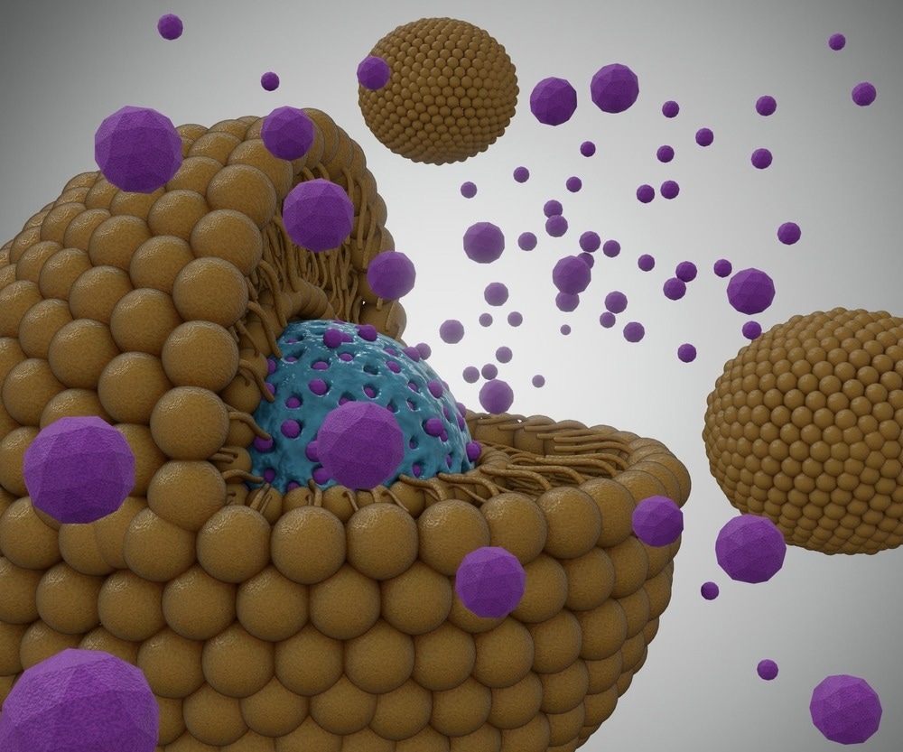
Image Credit: Love Employee/Shutterstock.com
TEM has shown that the most frequent uptake mechanism is endocytosis; nanocarriers have been reported interact with the plasma membrane and enter the cell encased in endosomes. This knowledge is beneficial in the design of effective delivery methods.
It can also be used in nanocarrier drug systems to examine the possible influence of biological surroundings on nanoformulations, including surface covering of most NPS by multilayered proteins known as the "protein corona."
The protein deposited on the NP surface is examined by TEM. Changes in NP parameters including aggregation, size, surface charge, mass, density, and absorbance are used to evaluate the adsorbed protein quantitatively. These observed values are then proportional to the quantity of protein adsorbed.
What Are the Challenges Associated with TEM?
TEM applications have certain limitations. When NPs are to be discovered inside organs ex-vivo, for example, an appropriate sampling can be achieved by removing numerous tiny tissue sections needed for TEM investigation from various organ parts.
Taking samples at various periods following NP administration allows for dynamic evidence of their uptake, relocation, and removal, while the tomographic technique allows for accurate 3D reconstructions of their positioning inside cells and tissues.
The main drawback of any high-resolution imaging technology is that only a small area of the specimen can be examined. Another problem is the requirement for frozen and divided samples: no live cells and no temporal resolution.
Recent Research
Petrushevska et al. demonstrated a new applicability of the TEM method for NP characterization in drug delivery systems. In this study, hybrid-silica xerogel particles were created as budesonide (BDS) carriers for effective local therapy of inflammatory bowel disease.
The TEM technique was utilized to check the long-term stability of the radiolabeled nanoparticles. It was also used to examine the shape, size and morphology of the particles of inorganic silica and hybrid silica that were developed.
The work was also aimed to generate self-assembled micelles of polymers for targeted delivery in tumor cells. TEM was also employed to evaluate the surface and uni-modal distribution of the particles.
Other Studies
Authors have used TEM to explore the structural properties of nanomaterials and biological specimens.
Sadauskas et al. used TEM to investigate the removal of gold nanoparticles (NPs) from mouse liver. Gold NPs were visualized histochemically in the liver using autometallographic labeling, and the accumulation of nanoparticles inside macrophage vesicular lysosome/endosome-like complexes was visualized using TEM.
Zhou et al. used TEM to investigate the production of drug-loaded, magnetic, hollow silica nanocomposites (MHSNC) coated with iron oxide magnetic nanoparticles (NPs) (10 nm) and silica on nanosized spherical and nano-needle like calcium carbonate.
The TEM results revealed that a smooth MHSNC surface and a thin layer of silica (10 nm) embedded with magnetic NPs had been successfully generated, and the nano-templates seemed to be dissolved.
Future Outlook
Nanomedicine research has advanced significantly as a result of the use of imaging tools, which are frequently integrated with multimodal approaches. Each technique has pros and cons and can only supply a limited amount of information.
The TEM technique is extremely useful for characterization of nanostructures. Furthermore, the requirement for ultrastructural analysis emphasizes the utility of TEM in the area of nanomedicine for drug delivery and targeting.
The long established TEM has only been partially utilized and provides researchers with extremely detailed images of materials at the microscopic and nano scale.
References and Further Reading
Malatesta, M. (2021). Transmission Electron Microscopy as a Powerful Tool to Investigate the Interaction of Nanoparticles with Subcellular Structures. International Journal of Molecular Sciences, 22(23), p. 22. doi.org/10.3390/IJMS222312789.
Zhou, W., et al., (2005). Drug-loaded, magnetic, hollow silica nanocomposites for nanomedicine. Nanomedicine: Nanotechnology, Biology and Medicine, vol. 1, no. 3, pp. 233–237. doi.org/10.1016/J.NANO.2005.06.005.
Petrushevska, M., et al. (2019). Transmission Electron Microscopy: Novel Application of Established Technique in Characterization of Nanoparticles as Drug Delivery Systems. PRILOZI, 40(2), pp. 67–72. doi.org/10.2478/PRILOZI-2019-0016.
Mahmood, S., et al. (2017). Advanced characterizations of nanoparticles for drug delivery: investigating their properties through the techniques used in their evaluations. Nanotechnology Reviews, 6(4), pp. 355–372. doi.org/10.1515/ntrev-2016-0050.
Berbel Manaia, E., et al. (2017). Physicochemical characterization of drug nanocarriers. International Journal of Nanomedicine, 12, pp. 4991–5011. doi.org/10.2147/IJN.S133832.
Vanhecke, D., et al. (2014). Quantification of nanoparticles at the single-cell level: an overview about state-of-the-art techniques and their limitations. Nanomedicine, 9(12), pp. 1885–1900. doi.org/10.2217/NNM.14.108.
Kola-Mustapha, A.T. (2019). Microscopy of Nanomaterial for Drug Delivery. Characterization and Biology of Nanomaterials for Drug Delivery, pp. 265–280. doi.org/10.1016/B978-0-12-814031-4.00010-6
Disclaimer: The views expressed here are those of the author expressed in their private capacity and do not necessarily represent the views of AZoM.com Limited T/A AZoNetwork the owner and operator of this website. This disclaimer forms part of the Terms and conditions of use of this website.