Quantum dots are molecular-sized nanocrystals that offer prospects for bioimaging due to their unique properties. This article focuses on how quantum dots can be used in zebrafish imaging applications.
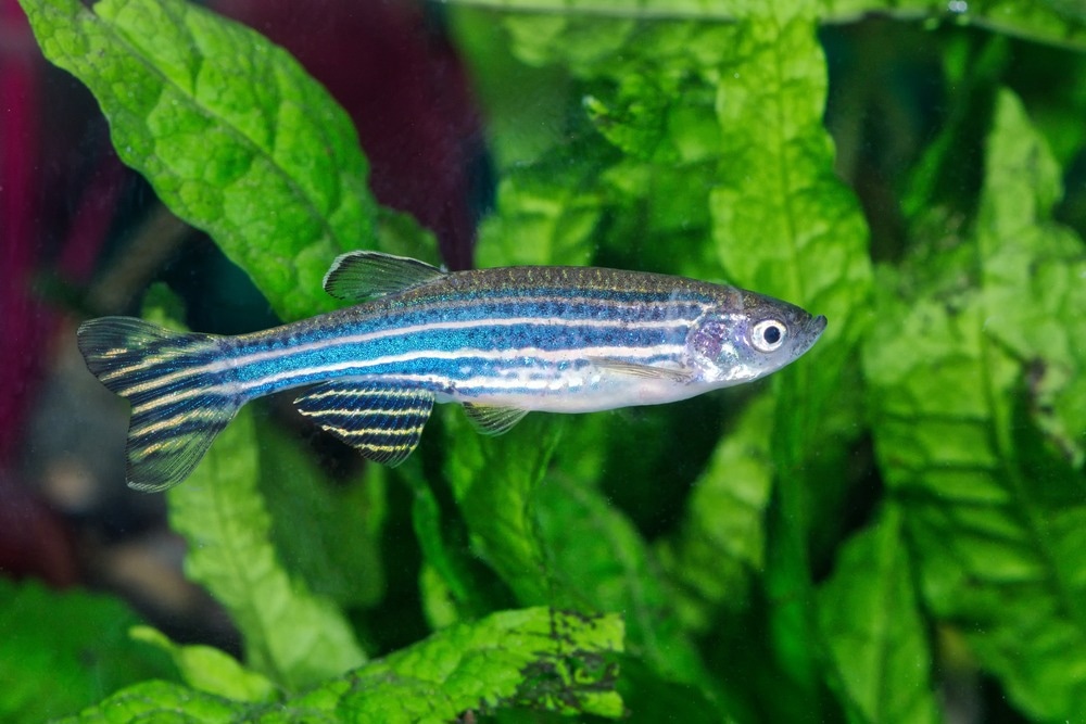
Image Credit: NERYXCOM/Shutterstock.com
Zebrafish Model and Imaging
The zebrafish organism has been utilized as a model to analyze biomaterials in various applications, such as bioimaging, simulating human disease, nanotoxicity, epigenomics, regenerative medicine, and DNA gene therapy.
It matches 69% of human genetic makeup, molecular structure, organ physiology, and cell growth. Consequently, it can serve as an alternative research model to humans. Key advantages of the model include short generation times, easy handling of zebrafish and embryos, and rapid embryonic development.
Embryonic development is crucial for examining the in-vivo system damage, toxicity, transport, and biocompatibility of nanoparticles and drugs. These developmental outcomes are used to flag any potential threats to human health and the environment.
The effectiveness of medical imaging tools can be assessed by the zebrafish live animal model with the incorporation of a nanomaterial.
For example, prior evaluation is necessary before using X-ray-based imaging techniques to scan human organs because X-ray exposure can damage cells due to its high energy. Zebrafish can be utilized to assess and fine-tune the X-ray energy intensity for human imaging applications.
Benefits of Using Quantum Dots for Zebrafish Imaging
Quantum dots are the ideal nanomaterial for imaging due to their high biocompatibility, ease of synthesis, minimal toxicity, and unique optical properties.
They also exhibit other notable advantages over the majority of organic fluorophore dyes, including size and wavelength-dependent luminescence, minimal photo-bleaching, fluorescence, narrow emission spectrum, increased brightness, photostability and large stokes shift.
Quantum dot-based imaging incorporates these properties to enhance fluorescence imaging providing benefits such as accurate and real-time in-vivo observations with high sensitivity, low costs, rapid responses, and no radiation.
Research on Quantum Dots in Zebrafish Imaging Applications
Multiphoton laser scanning microscopy (MPLSM), which produces several signals for image processing, is one imaging technique that can view embryos in three dimensions (3D+t) throughout time.
MPLSM involves second-harmonic generation (SHG), third-harmonic generation (THG), and two-photon-excited fluorescence. Light sheet fluorescence microscopy (LSFM) is another method utilized in 3D+t imaging.
A study published in the journal Chinese Physics B reported the use of zinc selenide (ZnSe) quantum dots for zebrafish imaging and two-photon fluorescence imaging. The employment of ZnSe quantum dots in biological imaging proved to have excellent biocompatibility.
ZnSe quantum dots displayed a two-photon cross-section of 9.1 × 10⁵ GM (1 GM = 10−50 cm4⋅s/photon) and after 24 hours of incubating zebrafish larvae with quantum dots, the fluorescence intensity of the ultra-violet (UV) light activated yolk sac was recorded to be 2.9 times larger than that of the control group.
Recent findings of novel "giant" core-shell quantum dots exhibiting record-breaking emissive lifetimes were highlighted in a publication of American Chemical Society's journal Nano Letters.
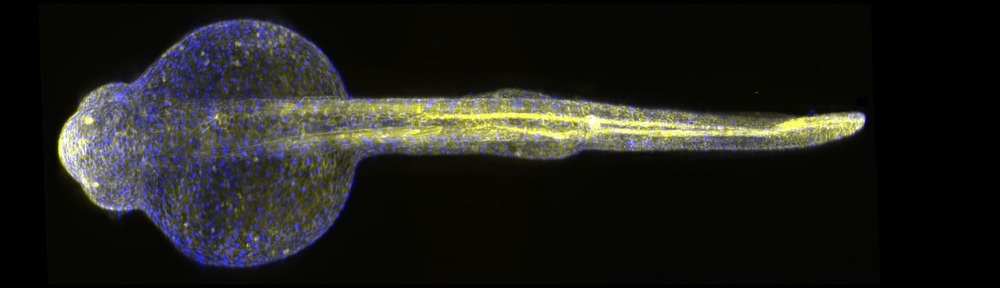
Image Credit: Micha Weber/Shutterstock.com
It is based upon the ability to manipulate the kinetic energy of the charge carrier on a parabolic potential energy surface to spatially localize holes or electrons within a shell/core heterostructure. This enables the use of cutting-edge techniques like time-gated single-particle imaging.
Researchers have used liquid metal jet X-Ray sources for high-resolution bio-imaging centered on an in-vivo zebrafish model; multi-functional green fluorescence carbon dots as a fluorescent probe to measure mercury, and laboratory-based X-ray NanoCT to analyze the morphological anatomy of embryos.
Kang et al., reported the successful application of carbon quantum dots (C-QDs) as an imaging probe. The dispersion (absorption, distribution, metabolism and excretion (ADME)) of C-QDs in zebrafish embryos and larvae was determined from their fluorescence emission, and zebrafish was used as an organism model to investigate the biotoxicity of C-QDs.
The results indicated that C-QDs have low toxicity and great biocompatibility as an imaging probe, and they do not affect the zebrafish embryo's ability to develop.
Graphene Quantum Dots (GQDs) have become an appealing candidate for two-photon fluorescence imaging (TPFI) due to their large two-photon absorption (TPA) cross-section and remarkable photostability.
Singh et al., reported in-vitro and in-vivo TPFI utilizing hydrothermally synthesized GQDs from Azadirachta indica extract. The ability of GQDs to act as a probe for two-photon fluorescence imaging of the digestive system was confirmed by in-vivo two-photon microscopy of zebrafish larvae and embryos.
This technique revealed high fluorescence of graphene quantum dots in the yolk sac area compared to other tissues.
Quantum dots have also been utilized as a microangiography contrast agent to visualize the cardiovascular system via zebrafish imaging to investigate their potential in skin research.
Commercial Example
Merck, an American multinational pharmaceutical corporation, has commercially available quantum dots for the purpose of bioimaging.
They manufacture products such as alloyed quantum dots, core-type quantum dots, and quantum dots with a core-shell. For example, core-shell quantum dots have CdSe in the core and ZnS in the shell.
Future Outlook
The synthesis of quantum dots, zebrafish rearing, embryo extraction, and administration of QDs into embryos and larvae are all covered in the literature reports on fluorescence imaging and bio-imaging of zebrafish, wherein nano-based quantum dots are used as imaging probes.
Novel quantum dots dramatically improve the zebrafish model's ability to detect the temporal dynamics of protein-protein interactions in living cells and expand research horizons for further studies.
Zebrafish imaging exploiting quantum dots as a probe is the finest illustration that can be applied in research, genetics, drug screening, and toxicity testing. In the near future, studies could focus on unraveling the genetics of human disease and discovering new treatments.
The market and scientific value of these nano-products have expanded as a result of recent advances in nanotechnology. Improvements in zebrafish embryo testing will therefore help researchers to further investigate the safety of pharmaceutical products for biomedical applications.
References and Further Reading
Pálmai, M., Beckwith, J. S., Emerson, N. T., Zhao, T., Kim, E. B., Yin, S., Parajuli, P., Tomczak, K., Wang, K., Sapkota, B., Tien, M., Jiang, N., Klie, R. F., Yang, H., & Snee, P. T. (2022). Parabolic Potential Surfaces Localize Charge Carriers in Nonblinking Long-Lifetime “giant” Colloidal Quantum Dots. Nano Letters. https://doi.org/10.1021/acs.nanolett.2c03563
Umar, E., Ikram, M., Haider, J., Nabgan, W., Haider, A., Imran, M., & Nazir, G. (2023). A state-of-the-art review on carbon quantum dots: Prospective, advances, zebrafish biocompatibility and bioimaging in vivo and bibliometric analysis. Sustainable Materials and Technologies, 35, e00529. https://doi.org/10.1016/J.SUSMAT.2022.E00529
Younis, M. R., He, G., Lin, J., & Huang, P. (2020). Recent Advances on Graphene Quantum Dots for Bioimaging Applications. Frontiers in Chemistry, 8, 424. https://www.frontiersin.org/articles/10.3389/fchem.2020.00424/full
Zhang, N.-N., 张楠楠, Zhou, L.-Y., 周立亚, Liu, X., 刘潇, Wei, Z.-C., 韦中超, Liu, H.-Y., 刘海英, Lan, S., 兰胜, Meng, Z., 孟钊, Fan, H.-H., & 范海华. (2021). Zebrafish imaging and two-photon fluorescence imaging using ZnSe quantum dots*. Chinese Physics B, 30(4), 044204. https://doi.org/10.1088/1674-1056/ABD467
Disclaimer: The views expressed here are those of the author expressed in their private capacity and do not necessarily represent the views of AZoM.com Limited T/A AZoNetwork the owner and operator of this website. This disclaimer forms part of the Terms and conditions of use of this website.