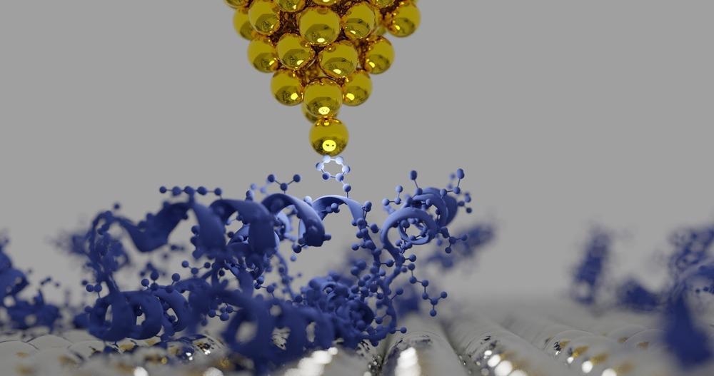
Atomic force microscopy (AFM) is an advanced microscopy technique that enables researchers to characterize the surface features of nanoparticles as small as 6 nm across.

Image Credit: sanjaya viraj bandara/Shutterstock.com
While it can be used as the sole experimental technique for nanoparticle research, AFM is often combined with other approaches.
What is AFM?
AFM is used to make 3D images at high levels of magnification down to a few nanometers. The technique was developed by IBM researchers Gerard Binning and Heinrich Rohrer in 1986.
AFM measures the interacting forces between the surface of the sample and an ultra-sharp probe. The probe is attached to a cantilever, which is attached to a laser-based sensor. Imaging software translates the sensor data into high magnification images.
AFM can be contact (where the probe touches the sample), non-contact (the probe is held just above the sample’s surface), or oscillating (the probe is made to resonate, and it contacts the surface at regular intervals). Due to its higher sensitivity, oscillating AFM is generally used for nanoparticle research.
AFM’s Advantages for Nanoparticle Research
AFM is advantageous in nanoparticle research because no surface modification or coating is needed for imaging. This has enabled scientists to perform topological analyses of small nanoparticles under 6 nm, such as ion-doped yttrium oxide (Y2O3), without any prior treatment.
Electron microscopy can usually not characterize low-density nanomaterials due to the poor contrast they present. However, AFM can provide topological analyses of these as well.
AFM has been used to characterize metal substrates based on nanoparticles used in sensors for surface-enhanced Raman spectroscopy (SERS).
The use of AFM led to a detection limit of barely one single molecule, enabling researchers to correlate the size, shape, and surface properties of different nanoparticles with enhanced SERS performance.
The main benefit of AFM for nanoparticle research is its ability to make 3D image scans. Other techniques are often incapable of characterizing the height or z-axis of nanoparticles.
The technique is also considerably less expensive, and devices take up less laboratory space than scanning electron microscopy (SEM) or transmission electron microscopy (TEM). However, AFM has slower scanning times than any kind of electron microscopy.
AFM at its Best in Combination with Other Techniques
In many cases, nanoparticle researchers combine multiple advanced characterization and analysis techniques to study multifaceted structures and events at the nanoscale.
Nanoscale materials display unexpected properties due to their extraordinarily high surface-to-volume ratio and the high molecular reactivity that comes with them.
A wide array of metrological techniques is used to discover the unexpected electronic, optical, chemical, and mechanical properties of these novel materials.
AFM has been combined with high-resolution TEM to tackle long-standing research gaps in nanotechnology, for example, to characterize the dendrimer template’s role in the growth of platinum nanoparticles. This combination also enabled the characterization of rhodium nanoparticles’ catalytic activity in phenylacetylene polymerization.
Kelvin probe force microscopy (KPFM) is an adapted form of non-contact AFM used to determine the work function of a sample’s surface. Work function is a property of a materials’ surface nanostructure, and relates to electron loss.
AFM and KPFM have been combined to make 3D maps of nanoparticles’ surface potential distributions, characterizing rust on steel and iron nanoparticles.
X-ray diffraction (XRD) is a commonly used technique for nanoparticle characterization. XRD is used to find out about nanoparticles’ crystalline structures, phase information, lattice parameters, and crystalline grain sizes.
Together, XRD and AFM have been employed to characterize silver nanoparticle thin films. The two techniques provide complementary information, especially the grain size and nanoparticle coverage data that AFM can provide combined with XRD’s ability to identify the preferential growth direction of the nanoparticles.
Another method that relies on X-ray technology is small-angle X-ray scattering (SAXS). SAXS is used to find out about nanoparticles’ size, size distribution throughout the sample, and shape.
Grazing-incidence SAXS (GISAXS) is a unique technique that detects diffusely scattered X-ray intensity from nanoscale objects and uses this to get information on nanoparticles.
AFM and GISAXS complement each other as well, with GISAXS providing localized morphological images of the surface and AFM backing this up with 3D topographical data. The two methods have been used to study silicon dots produced by ion bombardment, as well as self-assembled iron oxide nanoparticles.
Using AFM to Design New Nanoparticles
Researchers have also used AFM to help with the design of new carbon-based nanocomposites.
AFM was used to identify the structure-property relationship in a new compound. This kind of analysis is important in the development of new nanomaterials, and also drugs and pharmaceuticals.
AFM can provide information about the dispersion and morphological and topographical features of resins loaded with various nanoparticles, including the carbon nanostructured doping agents studied in this research.
The technique is adopted for its non-destructive, non-contact capabilities, which have minimal effect on the structure-property relationship of novel nanoparticles and other materials.
Continue reading: Park FX40: The Story Behind Designing a New Class of the Automatic Atomic Force Microscope (AFM).
References and Further Reading
Mourdikoudis, S., R. M. Pallares, and N. T. K. Thanh (2018) Characterization techniques for nanoparticles: comparison and complementarity upon studying nanoparticle properties. Nanoscale. Available at:https://pubs.rsc.org/en/content/articlelanding/2018/NR/C8NR02278J
Raimondo, R. et al. (2019) Carbon-Based Aeronautical Epoxy Nanocomposites: Effectiveness of Atomic Force Microscopy (AFM) in Investigating the Dispersion of Different Carbonaceous Nanoparticles. Polymers. Available at: https://www.mdpi.com/2073-4360/11/5/832
Disclaimer: The views expressed here are those of the author expressed in their private capacity and do not necessarily represent the views of AZoM.com Limited T/A AZoNetwork the owner and operator of this website. This disclaimer forms part of the Terms and conditions of use of this website.