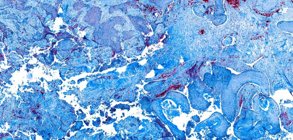
Oral cancers are amongst the most common types of cancer worldwide. Their incidence amongst younger individuals has increased notably in recent decades with little change in mortality compared with other cancers such as breast cancer.

Image Credit: Carl Dupont/Shutterstock.com
Surgery and radiotherapy are frequently employed to combat oral cancers, but as these techniques can leave aesthetically displeasing scarring and muscular defects with implications for the patient's mental health, there is a call for alternatives.
Nanomaterials are defined as having a dimension on the nanoscale, with particles constructed from gold for biomedical applications typically in the 5 – 100 nm range. Gold nanoparticles have attracted particular attention as cancer therapeutic and diagnostic agents and theranostics, owing to their unique properties and applicability to delivery applications.
How are Gold Nanoparticles Useful in Cancer Diagnosis?
Nanoscale metallic gold engages in a phenomenon known as localized surface plasmon resonance, wherein adsorption is enhanced significantly at a specific wavelength of light related to the size and shape of the particle. The surface plasmon resonance peak wavelength is highly tunable throughout the visible and near-infrared region by controlling particle morphology. Gold nanoparticles have applications as colorimetric sensors and diagnostic probes as a result.
Gold nanoparticles are already commonly used in devices such as pregnancy tests and COVID-19 lateral flow tests as colorimetric indicators. Here, a ligand complementary to the target molecule is applied to the nanoparticle's surface via a coating. When the target is present, the nanoparticles will stick to an indicative line or aggregate and produce a color change.
In the context of oral cancer diagnostics, several gold nanoparticle probes have been developed that bind with oral cancer biomarkers in saliva and can indicate their presence by a slight change in surface plasmon resonance wavelength caused by the change in dielectric constant surrounding the particle.
Gold nanoparticles can also be employed as the substrate in surface-enhanced Raman spectroscopy experiments, which can quantitatively establish the presence of biomarkers bound to the particle after mixing with saliva with extremely low detection limits.
For example, oral squamous cell carcinoma cells are widely reported to overexpress epidermal growth factor receptors EGFR, ErbB1, and HER1. Gold nanoparticles bound with anti-epidermal growth factor receptor have been mixed with saliva to allow the biomarker to bind, and then separated and analyzed by SERS.
Due to the disordered genome of cancer cells, they often overexpress one or more receptors on their exterior surface, and thus the complementary ligand to an overexpressed receptor can be bound to the gold nanoparticle to impart active targeting functionality. As already discussed, the surface plasmon resonance of gold nanoparticles can be tuned into the near-infrared region, which is the wavelength of light most penetrating through biological tissue, being absorbed less than shorter wavelength visible light by hemoglobin and water molecules in the body.
In combination with the targeting specificity of gold nanoparticles, this feature can be exploited to clearly define the volume of the tumor for diagnostic purposes. Further, the high density of metallic gold presents applications as X-ray, ultrasound, and computed tomography contrast agents.
How are Gold Nanoparticles Useful in Cancer Therapy?
Surface plasmon resonance in gold nanoparticles is a result of in-phase oscillations of the electron cloud surrounding the particle with incident light.
When in-phase with oscillating electrons, incident photons are absorbed, producing the observed color of gold nanoparticle colloids. Thus, when high-energy monochromatic incident light of the surface plasmon resonance wavelength is applied to gold nanoparticles, the photons impart rapid heating by absorption capable of inducing localized hyperthermia.
Gold nanoparticle-assisted photothermal therapy has demonstrated great potential in destroying tumors in a highly specific manner, where heating cancer cells just a few degrees Celsius can cause proteins to denature and induce apoptosis.
Higher energy X-ray or gamma-ray lasers employed in radiotherapy can also interact with the electron cloud of gold nanoparticles and initiate a process known as an Auger cascade, wherein an incident photon can cause the ejection of an inner shell electron and prompt another to fall to the now vacant lower energy level.
Upon falling to a lower energy level, a photon is released bearing an energy equivalent to the difference in energy between the electron orbitals, which may go on to interact with another electron and cause its ejection.
Auger cascades of as many as ten electrons from a single gold nanoparticle have been documented, and the released electrons can go on to either damage biological targets directly or otherwise interact with surrounding water or oxygen molecules to generate reactive oxygen species that go on to damage the cell.
In this way, gold nanoparticles make potent radiation dose enhancers that can be firstly localized to the tumor site by active targeting mechanisms, used to precisely identify the tumor's dimensions, and then engage in localized cell destruction.
The great customizability of the gold nanoparticle surface also allows a wide range of chemotherapeutic drugs to be delivered to the tumor alone or simultaneously with a payload of synergistic drugs in a highly specific manner. Doing so enhances their efficacy and reduces off-target effects.
Importantly, the drug delivery functionality of gold nanoparticles can be combined with photothermal or radiotherapy in a combined approach, taking advantage of all the unique properties of the nanomaterial. Rapid heating can also be utilized as a mechanism of triggered release for the drug payload, ensuring that the cytotoxic cargo is only released in the vicinity of the tumor. Currently, no gold nanomaterial is engaged in a clinical trial specifically for the treatment of oral cancer, though several are engaged in trials for other specific cancers and will eventually be translated over.
References and Further Reading
Zhang, Q., et al. (2022) Gold nanomaterials for oral cancer diagnosis and therapy: Advances, challenges, and prospects. Materials Today Bio. https://www.sciencedirect.com/science/article/pii/S2590006422001314
Kimura, I., Kitahara, H., Ooi, K., Kato, K., Noguchi, N., Yoshizawa, K., Nakamura, H. & Kawashiri, S. (2016). Loss of epidermal growth factor receptor expression in oral squamous cell carcinoma is associated with invasiveness and epithelial-mesenchymal transition. Oncology Letters, 11 (1).https://www.ncbi.nlm.nih.gov/pmc/articles/PMC4727181/
Kah, et al. (2007) Early diagnosis of oral cancer based on the surface plasmon resonance of gold nanoparticles. International journal of Nanomedicine, 2(4). https://pubmed.ncbi.nlm.nih.gov/18203445/
Disclaimer: The views expressed here are those of the author expressed in their private capacity and do not necessarily represent the views of AZoM.com Limited T/A AZoNetwork the owner and operator of this website. This disclaimer forms part of the Terms and conditions of use of this website.