Article Updated on 17 May 2021
The world of the nanomolecular is an enigmatic one. To understand the physical, chemical, and nuclear properties that shape the world, scientists have devised ever-more sophisticated methods over the last century. However, several classes of functional materials that have emerged in recent years have atomic structures limited to the nanoscale. These include nanoparticles, nanostructured materials, nanocomposites and bulk crystals which have an intrinsic nanoscale order.

Image Credit: Isuaneye/Shutterstock.com
Working at the Nanoscale
Aside from these emerging materials, several structures in nature exist at the nanoscale. Hemoglobin has a diameter of 5.5 nm, for example. Naturally occurring nanoparticles exist in smoke from fires and in volcanic ash. The nanoscale covers a range of dimensions from 1-100 nm.
Working at the nanoscale requires an understanding of the various dimensions and types of nanomaterials, as well as the atomic interactions which cause the physical and chemical properties that these materials and substances display and how to manipulate them. Currently, there is no “hard and fast” solution to the problems of analyzing the atomic structure of substances at the nanoscale.
One method that researchers can employ to carry out this task is crystallography. However, this reliable technique has issues when working at such minuscule scales.
What is Crystallography?
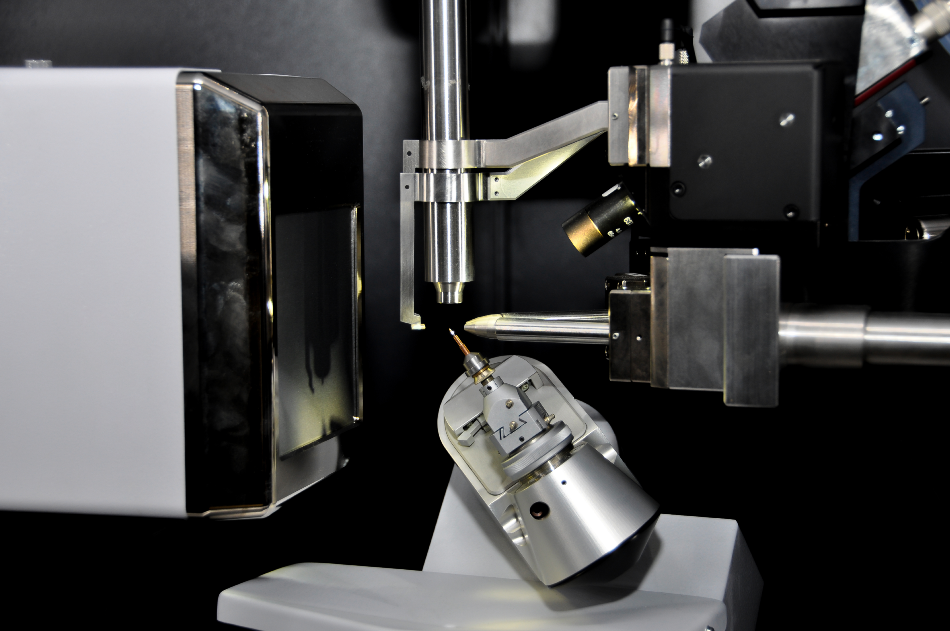
Crystallography is a field of science concerning the analysis of atomic structures in crystalline solids using a specific set of analytical techniques. By using the known properties and inner structures of crystals, a crystallographer can determine the arrangement of atoms, which generates knowledge about the chemical and physical properties of the substance.
Analogous to microscopy, crystallography involves firing a beam at a sample to create an image of the structure. However, as light has a diffraction limit greater than the scale needed for analysis of these structures, other forms of radiation are used in this technique.
Solid materials are characterized by crystallography using X-ray, neutron, and electron diffraction techniques. Many other analytical techniques provide information that is incorporated into the field of crystallography, including spectroscopic techniques, X-ray fluorescence, and computer modeling and visualization. To study a substance via crystallographic techniques, first, a stable crystal must be created: various methods are employed by crystallographers and some substances are harder to crystallize than others.
Crystallography has been one of the driving forces behind major advances in fields such as synthetic chemistry and genetics and has contributed to the development of several drugs that are in use today.
Overcoming the Problems of Crystallography at the Nanoscale
The main problem with crystallographic studies at the nanoscale concerns growing crystals at such small scales. Many methods have been proposed to overcome this issue.
One promising study carried out by scientists working at Imperial College London involved the creation of a “baking tray” for forming stable crystals at the nanoscale. By attaching polymers to graphene and creating small pockets on the graphene analogous to a baking tray, proteins were attracted to the grafted molecules, making it easier to nucleate a stable crystal that could grow large enough to analyze with X-ray crystallographic methods.
Another promising method for studying details at the nanomolecular scale using crystallography techniques is fluctuation X-ray scattering. First proposed more than 40 years ago, this technique uses ultrashort pulses of X-ray radiation which are scattered by macromolecules in solution. This avoids motion blur (which comes about from the rapid spin and movement of proteins) and yields a higher degree of structural information.
Fluctuation scattering uses data from many particles at once, which means an increase in efficiency as only a few minutes are needed to perform the analysis, rather than the several hours or days required by more conventional methods. This method has been used to study the PBCV-1 virus in far greater detail than before.
Another promising method in the field of metal nanocrystals is the seed-mediated growth method, where nucleation and growth are separated in a 2-step process, using a seed particle to grow a crystal in a solution including metal precursors, reducing reagents, and additives that tune the shape of the crystal produced.
To perform accurate crystallographic studies at the nanoscale, it has been proposed that researchers from many fields including materials science, computer science, synthetic chemistry, and applied mathematics will have to collaborate, working within a “complex modeling” paradigm. Other analytical and imaging techniques that are proving useful include TEM (Transmission Electron Microscopy.)
This emerging field of research is an interesting one and represents a challenge to crystallography as a method of atomic and structural analysis. As more powerful crystallography machinery, more effective methods, and the increased cross-over with other scientific disciplines are developed, the world of nanomolecular using crystallography techniques will become easier than ever before.
Sources
Billinge, S.J.L. and Levin, I. (2007) The Problem with Determining Atomic Structure at the Nanoscale, Science Vol. 316, Issue 5824, Pgs. 561-565
https://science.sciencemag.org/content/316/5824/561
Working at the Nanoscale, Nano.Gov
https://www.nano.gov/nanotech-101/what/working-nanoscale
Liz-Marzán, L.M. and Grzelczak, M. (2017) Growing anisotropic crystals at the nanoscale, Science Vol. 356, Issue 6343, Pgs. 1120-1121
https://science.sciencemag.org/content/356/6343/1120
Govada, L. et al. (2016) Exploring Carbon Nanomaterial Diversity for Nucleation of Protein Crystals Nature Sci. Rep 6, article 20053
https://www.nature.com/articles/srep20053
Niu, W. et al. (2013) Seed-mediated growth of noble metal nanocrystals: crystal growth and shape control Nanoscale Issue 8
https://pubs.rsc.org/en/content/articlelanding/2013/nr/c3nr00219e
Pande, K et al. (2018) Ab initio structure determination from experimental fluctuation X-ray scattering data PNAS Issue 115 Vol. 46 11772-11777
https://www.pnas.org/content/115/46/11772
Disclaimer: The views expressed here are those of the author expressed in their private capacity and do not necessarily represent the views of AZoM.com Limited T/A AZoNetwork the owner and operator of this website. This disclaimer forms part of the Terms and conditions of use of this website.