The family of electron microscopy techniques have become staple methods for imaging nanoscale objects, including nanoparticles, viruses and proteins. The appeal of electron microscopy as a characterization and research technique is the very high spatial resolutions achievable with electron microscopes. Sub-Angstrom resolutions are now routinely achievable, and it is possible to record three-dimensional structures as well as two-dimensional images as well.
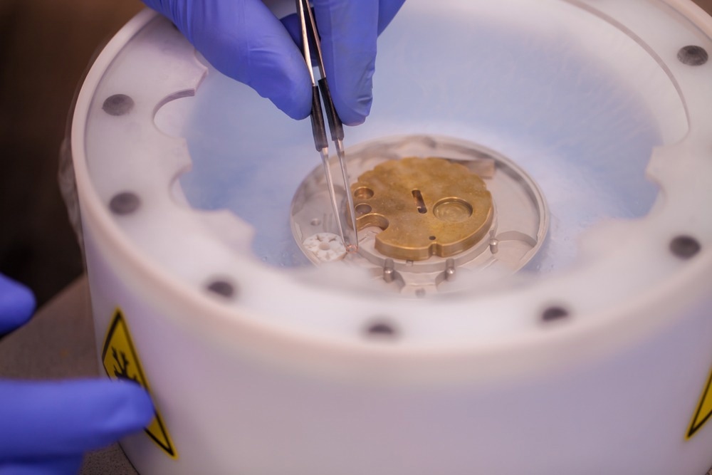
Image Credit: PolakPhoto/Shutterstock.com
An electron microscope consists of an electron gun, various lenses and optics, a sample holder and detectors oriented either in a transmission or scattering geometry. Due to the high voltages often applied to electron guns, there is a significant risk of electrical discharges or arcs within the apparatus. One approach to minimizing this risk is to keep the experiment under high vacuum conditions.
The use of high vacuum conditions poses some challenges for the sample preparation and the types of samples that can be used within the electron microscope. Another issue that needs to be accounted for in the sample preparation for electron microscopy is the effects of electron beam damage on the sample. Exposure to high-intensity electron beams can cause radiation damage and degradation of the sample, which can be detrimental to the quality of the images obtained.
The right sample preparation can help minimize radiation damage and help ensure surfaces are clean and free from contamination, which may obscure features or complicate elemental analysis.
Sample Preparation
What type of sample preparation method is required depends on whether measurements are to be made using transmission electron microscopy or scanning electron microscopy methods. Transmission measurements generally require thinner samples than measurements that use the secondary electrons emitted from the sample as in transmission mode, the electron beam is directly imaged having passed through the sample and so must not be completely attenuated by the sample.
Sample preparation methods are also dependent on the material type to be imaged. Nanoparticles are usually dispersed onto transmission electron microscopy grids in a solvent matrix. The solvent matrix is then removed using methods like air drying.
Some care needs to be taken to avoid drying artifacts, particularly with nanoparticles, as the nanoparticles can aggregate during drying, and so the measured image is not of their in situ colloidal dispersion state.
Transmission measurements on metals are relatively straightforward. A thin disc of material is prepared and then polished or milled until the surfaces are clean of contaminants and suitable for imaging. Sometimes methods such as focused ion beam or ion milling are used to achieve the very high quality and high surface evenness required to take good images.
Larger samples are sometimes sectioned and cut into slices, then mounted on transmission electron microscopy grids for imaging.
Scanning Electron Microscopy
Scanning electron microscopy sample preparation requirements are generally less demanding than transmission electron microscopy, but larger samples may need to be cut or machined down to fit into sample holders or imaging errors. Surface smoothness can be important as unevenness on the surface can cause overlapping of the electron signals and obscure features of interest from the sample.
Epoxy impregnation is often used for measuring porous materials such as concretes. The epoxy sets into any cracks or holes in the structure, preventing any further cracks formed from sample drying when the sample loses moisture under the vacuum conditions for measurement or undergoes other sample preparation procedures.
Resins can also be used for the measurement of biological samples. There have been recent developments in the use of conductive resins for electron microscopy imaging. One of the challenges with using electrons as opposed to light for sample visualization is that there can be charging effects on the sample that reduce the effective resolution, introduce difficult-to-identify and understand artifacts and limit the intensity of electron beams that can be used.
The use of conductive materials can help reduce the effects of sample charging as all the electrons are absorbed and channeled to ground. Sometimes samples are intentionally coated in a highly conductive material such as gold to minimize charging effects.
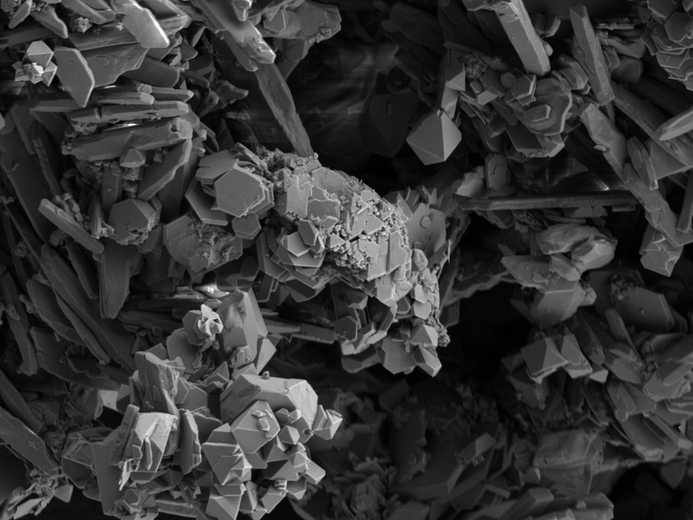
Image Credit: V/Shutterstock.com
Cryo-EM
Biological samples pose their own series of sample-handling challenges for electron microscopy methods. Many biological samples are fragile and easily damaged by exposure to the electron beam, have a high water content that is not compatible with a vacuum environment, and are also dynamic in their nature, so they undergo structural changes.
The development of cryogenic electron microscopy, known as cryo-EM, has revolutionized the possibilities for structural analysis of biological samples with electron microscopy. Cryo-EM uses very specific sample preparation methods where samples are flash-frozen using cryogenic liquids.
There are several stages to sample preparation for cryo-EM, including steps such as adding buffers or changing pH conditions to improve sample stability and using different sample carriers, deposition and vitrification methods. The right choice at each step is often highly sample-dependent and requires some optimization to obtain the highest quality electron images.
Sample preparation is a critical step in obtaining the best results from electron microscopy measurements and can be as important as instrumentation set-up for achieving the full resolving capabilities of these techniques.
References and Further Reading
Ali, S., Ali, E., Abro, R., Saleem, F., Khan, A., Ahmed, I., Ahmed, M., Nizamuddin, S., Hussain, T., Hossain, N., Mujawar, N., & Shah, A. (2021) Nanomaterials : Applications , waste-handling , environmental toxicities , and future challenges – A review. Journal of Environmental Chemical Engineering, 9(2), p. 105028. https://doi.org/10.1016/j.jece.2021.105028
Robertson, M. J., Meyerowitz, J. G., & Skiniotis, G. (2022) Drug discovery in the era of cryo-electron microscopy. Trends in Biochemical Sciences, 47(2), pp. 124–135. https://doi.org/10.1016/j.tibs.2021.06.008
Subramaniam, S., & Milne, J. L. S. (2004) Three-dimensional Electron Microscopy at Molecular Resolution. Annu. Rev. Biophys. Biomol. Struct., 33, pp. 141–155. https://doi.org/10.1146/annurev.biophys.33.110502.140339
Baker, L. A., & Rubinstein, J. L. (2010) Radiation Damage in Electron Cryomicroscopy. In Methods in Enzymology (Vol. 481, Issue 10). Elsevier Masson SAS. https://doi.org/10.1016/S0076-6879(10)81015-8
Zhang, W., Melo, L. G. D. A., Hitchcock, A. P., & Bassim, N. (2019) Electron beam damage of epoxy resin films studied by scanning transmission. Micron, 120 (December 2018), pp. 74–79. https://doi.org/10.1016/j.micron.2019.02.003
Michen, B., Geers, C., Vachecke, D., Endes, C., Rothen-Ruithauser, B., Balog, S., & Petri-Fink, A. (2015) Avoiding drying-artifacts in transmission electron microscopy : Characterizing the size and colloidal state of nanoparticles. Scientific Reports, 5, p. 9793. https://doi.org/10.1038/srep09793
Sasaki, H., Matsuda, T., Kato, T., Muroga, T., Iijima, Y., Saitoh, T., ... & Hirayama, T. (2004) Specimen preparation for high-resolution transmission electron microscopy using focused ion beam and Ar ion milling. Microscopy, 53(5), pp. 497-500. https://doi.org/10.1093/jmicro/dfh067
Bisschop, J., & Mier, J. G. M. Van. (2002). How to study drying shrinkage microcracking in cement-based materials using optical and scanning electron microscopy ? Cement and Concrete Research, 32, pp. 279–287. https://doi.org/10.1016/S0008-8846(01)00671-8
Nguyen, H. B., Thai, T. Q., Saitoh, S., Wu, B., Saitoh, Y., & Shimo, S. (2016) Conductive resins improve charging and resolution of acquired images in electron microscopic volume imaging. Scientific Reports, 6, p. 23721. https://doi.org/10.1038/srep23721
Postek, M. T., & Vladár, A. E. (2015) Does Your SEM Really Tell the Truth ?— How Would You Know ? Part 4 : Charging and its Mitigation. Proc SPIE Int Soc Opt Eng, 9636, pp. 1–21. https://doi.org/10.1117/12.2195344.
Weissenberger, G., Henderikx, R. J. M., & Peters, P. J. (2021) Understanding the invisible hands of sample preparation for cryo-EM. Nature Methods, 18, pp. 463–471. https://doi.org/10.1038/s41592-021-01130-6
Disclaimer: The views expressed here are those of the author expressed in their private capacity and do not necessarily represent the views of AZoM.com Limited T/A AZoNetwork the owner and operator of this website. This disclaimer forms part of the Terms and conditions of use of this website.