Oct 5 2013
Scientists have developed a new kind of ‘X-ray vision’ that is able to peer inside an object and map the three-dimensional distribution of its nano-properties in real time.
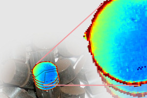 The new imaging technique can reveal the nano-properties of objects
University of Manchester researchers, working with colleagues in the UK, Europe and the US, say the novel imaging technique could have a wide range of applications across many disciplines, such as materials science, geology, environmental science and medical research.
“This new imaging method – termed Pair Distribution Function-Computed Tomography – represents one of the most significant developments in X-ray micro tomography for almost 30 years,” said Professor Robert Cernik in Manchester’s School of Materials.
“Using this method we are able to image objects in a non-invasive manner to reveal their physical and chemical nano-properties and relate these to their distribution in three-dimensional space at the micron scale.
“Such relationships are key to understanding the properties of materials and so could be used to look at in-situ chemical reactions, probe stress-strain gradients in manufactured components, distinguish between healthy and diseased tissue, identify minerals and oil-bearing rocks or identify illicit substances or contraband in luggage.”
The research, published in the journal Nature Communications, explains how the new imaging technique uses scattered X-rays to form a three-dimensional reconstruction of the image.
“When X-rays hit an object they are either transmitted, absorbed or scattered,” explained Professor Cernik. “Standard X-ray tomography works by collecting the transmitted beams, rotating the sample and mathematically reconstructing a 3D image of the object. This is only a density contrast image, but by a similar method using the scattered X-rays instead we can obtain information about the structure and chemistry of the object even if it has a nanocrystalline structure.
“By using this method we are able to build a much more detailed image of the object and, for the first time, separate the nanostructure signals from the different parts of a working device to see what the atoms are doing in each location, without dismantling the object.”