Films and coatings are used in a variety of applications, such as thin films for food protection, coatings on medical devices, and drug delivery coatings, etc, and hence make them essential for evaluation of these materials. Confocal Raman Microscopy is a unique tool that enables non-destructive imaging of the chemical composition of heterogeneous components within a coating or film. The confocal arrangement offers the highest spatial resolution and also helps in acquiring depth profiles that are suitable for the 3D characterization of polymer films and coatings sans sample preparation.
Confocal Raman Microscopy
WITec’s alpha300 R is an advanced Confocal Raman Microscope that integrates a high-transmission Raman spectroscopy system and a highly sensitive confocal microscope. With 200nm resolution at each image pixel, a complete spectrum is normally obtained in just 10 to 100ms. The number of spectra or image points is restricted only by the computer memory. A standard image includes 10,000 (100 x 100) to 65 536 (256 x 256) spectra. From this multi-spectrum file, an image is produced by combining over a particular Raman line or a region in all spectra.
Since all spectra are stored in memory, a range of properties such as peak position, peak-width, or center of mass of certain Raman lines can be measured from a single measurement. When measuring depth profile, the focal plane can be shifted in the z-direction when performing either xz scans or creating x-y image stacks in the z direction.
Polymer Coating
For the experiment, the inner polymer coating of an orange juice container was examined using the alpha300 R system. An x-z scan was performed with a scan range of 50 ìm x 100 ìm at 200 x 120 pixels (= 24 000 spectra) by means of a 100x objective (NA = 1.25). A 532nm Nd:Yag laser was utilized for excitation; the acquisition time for each spectrum was 50ms.
Results and Discussion
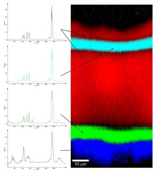
Figure 1. Raman spectra (left) and Raman image (right) of the inner coating of an orange juice container.
In Figure 1 (left), four separate spectra can be seen within the acquired multi-spectrum file. Each spectrum stands for a particular chemical compound within the polymer film. With the help of integrated software tools, the distribution of each compound can be observed by examining all acquired spectra. Figure 1 (right) depicts the resulting color-coded image, clearly showing that the coating includes five different layers even though only four components are involved. The software also helps in measuring distances within the image, signifying that all layers differ in thickness.
Analysis of Heterogeneous Layered Samples
The alpha300 R Confocal Raman Microscope from WITec is suitable for the analysis of heterogeneous layered samples on the sub-micrometer scale. As indicated in Figure 1, different polymer layers in the film can be easily resolved and correlated with their related Raman-Spectrum. Using the depth profiling capabilities of the Confocal Raman Microscope, complete information on the structure of multilayered polymer coatings or films can thus be obtained.
Conclusion
WITec’s alpha300 R Confocal Raman Microscope is an ideal tool for the 3D characterization of polymer films and coatings without sample preparation. WITec specializes in manufacturing high-performance instrumentation for industrial and scientific applications focused on innovative solutions for optical and scanning probe microscopy.

This information has been sourced, reviewed and adapted from materials provided by WITec GmbH.
For more information on this source, please visit WITec GmbH.