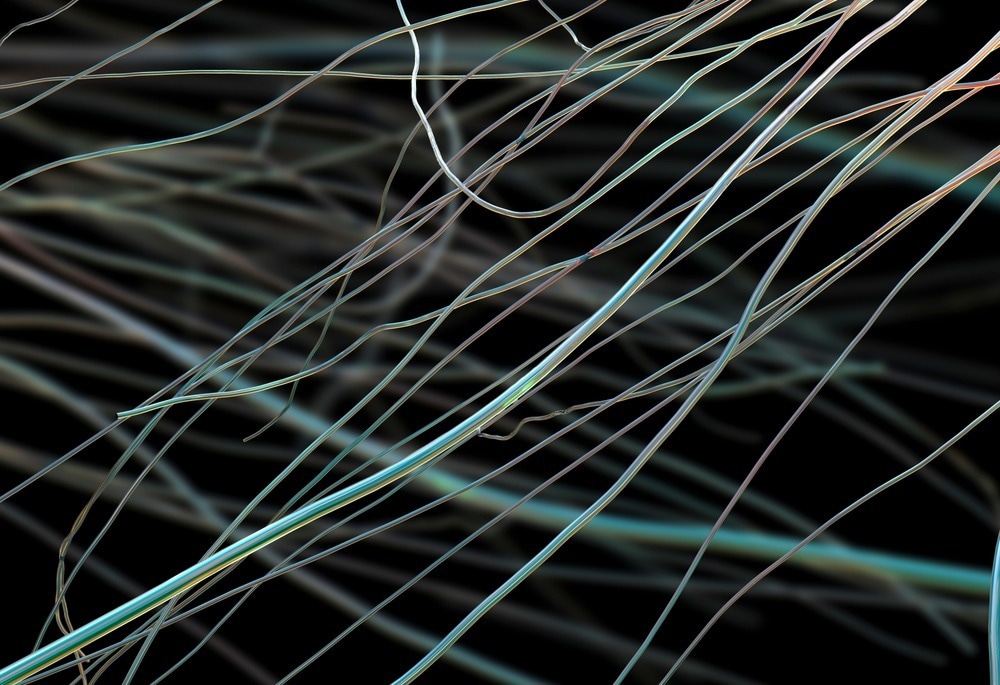
This article discusses the role of scanning electron microscopy (SEM) imaging in investigating the morphology and pore size distribution (PSD) of nanofibers.

Image Credit: Kateryna Kon/Shutterstock.com
Importance of Morphology and PSD Analysis in Nanofibers
Nanofibers have gained significant attention for different applications, such as barrier fabrics and high-efficiency filters. In biomedical applications, nanofibers have been used as drug delivery systems, scaffolds in wound dressings, and carriers for cell cultivation and for enzyme immobilization.
The internal structure of nanofibers makes them suitable as supporting materials in cell therapy, which is an effective treatment approach for treating different diseases, including corneal defects and chronic wounds. Similarly, nanofiber scaffolds support the seeded cells before in-vivo transplantation when used as cell cultivation carriers.
Scaffold requirements include controlled porosity, permeability, biocompatibility, and suitable mechanical properties comparable to natural tissue. Additionally, the morphology of the scaffolds plays a critical role in biomedical applications such as stem cell transplantation and wound dressing.
Specific surface area, size, and volume of the pores significantly affect cell growth, proliferation, and adhesion. Moreover, the release of incorporated biologically active substances is influenced by the internal architecture of the nanofibers. These factors increase the importance of morphology and PSD analysis of nanofibers.
Application of SEM for Nanofiber Morphology and PSD Investigation
Currently, imaging methods such as SEM are used extensively for the morphological characterization of several materials, including nanofibers, as they allow direct visualization of the observed nanostructures.
Thus, images obtained through SEM imaging provide crucial information required to compare the local structures within the entire nanofibrous sample, which is a significant advantage of the method.
Moreover, SEM can also evaluate in vitro biomedical experiments as it can depict the cell cultivation process on several synthetic substrates. However, a quantitative comparison among different nanofibrous materials cannot be performed using imaging techniques such as SEM as these techniques do not provide defined numerical values.
Thus, SEM can be used to investigate the primary characteristics of prepared nanofibers, such as fiber diameter. Additionally, the method can reveal heterogeneities in the nanofibrous structures that emerge during electrospinning under specific electrospun solution conductivity and polymer concentration.
The application of SEM is crucial to investigate nanofibrous materials with advanced structures, such as core-shell structures obtained by coaxial electrospinning. SEM has been used to evaluate the influence of electrospinning conditions on the formation of nanocoil structures.
SEM is also an essential method for evaluating the nanofiber orientation, which affects the mechanical properties of the nanofiber and plays a crucial role in biological applications.
In a study published in the Journal of Nanomaterials, researchers used the SEM method to determine the fiber diameters and identify the heterogeneities in nanofibrous structures. Nanofibers used in this study were prepared from polyamide, gelatin, poly(ε-caprolactone), and polylactide using the needleless electrospinning technique.
Mean values of 30 measurements on the SEM images were used to determine the fiber diameters at 5000x magnification. The thinnest fibers were obtained from gelatin with a mean value of 110 nm.
Heterogeneities were identified in nanofibers prepared from polylactide and poly(ε-caprolactone) under the initial electrospinning conditions. In both cases, optimization of the electrospinning conditions/process prevented the creation of these heterogeneities and facilitated the formation of homogenous structures.
Additionally, SEM analysis also revealed heterogeneities in the structure that can significantly impact the nanofibrous material behavior in medicinal applications, such as drug release and cell cultivation.
In another study published in the Textile Research Journal, researchers investigated the morphology and PSD of nanofiber membranes obtained from nylon 6 using force-spinning and electrospinning methods. All samples were sputter coated with gold before SEM analysis. Fiber diameter was measured using Image J on SEM micrographs.
SEM images of forcespun and electrospun nanofiber mats demonstrated that most fibers were in the sub-micron range. However, the electrospun samples displayed a more uniform and regular fiber morphology than forcespun samples, attributed to the differences in centrifugal force- and electrical field-driven fiber formation mechanisms.
Additionally, nano-nets with interlinked ultra-fine fibrils and diameters in the range of tens of nanometers were also observed in the electrospun nanofiber mats. Nano-net structures can significantly affect the barrier properties and PSD as these structures divide the voids between the coarser nanofibers.
The fiber diameter was impacted by the spinning solution viscosity for both spinning methods. Larger fiber diameters were observed in forcespun and electrospun membranes when they were prepared from 20 wt% polymer solution concentration. Both membrane areal density and fiber diameter significantly influenced the membrane PSD.
To summarize, the application of SEM imaging has become crucial for the observation of nanofibrous structures, specifically in biomedical applications. In the future, the growing usage of nanofibers in other applications will further increase the importance of SEM imaging due to the need for a complex approach in the morphological characterization of nanofibrous materials.
References and Further Reading
Sirc, J., Hobzova, R., Kostina, N.Y., Munzarová, M., Juklíčková, M., Lhotka, M., Kubinova, S., Zajicova, A., Michalek, J. (2012). Morphological Characterization of Nanofibers: Methods and Application in Practice. [Online] Journal of Nanomaterials. Available at: https://www.researchgate.net/publication/258383635_Morphological_Characterization_of_Nanofibers_Methods_and_Application_in_Practice
Krifa, M., Yuan, W. (2015). Morphology and pore size distribution of electrospun and centrifugal forcespun nylon 6 nanofiber membranes. [Online] Textile Research Journal. Available at: https://www.researchgate.net/publication/282904326_Morphology_and_pore_size_distribution_of_electrospun_and_centrifugal_forcespun_nylon_6_nanofiber_membranes
Disclaimer: The views expressed here are those of the author expressed in their private capacity and do not necessarily represent the views of AZoM.com Limited T/A AZoNetwork the owner and operator of this website. This disclaimer forms part of the Terms and conditions of use of this website.