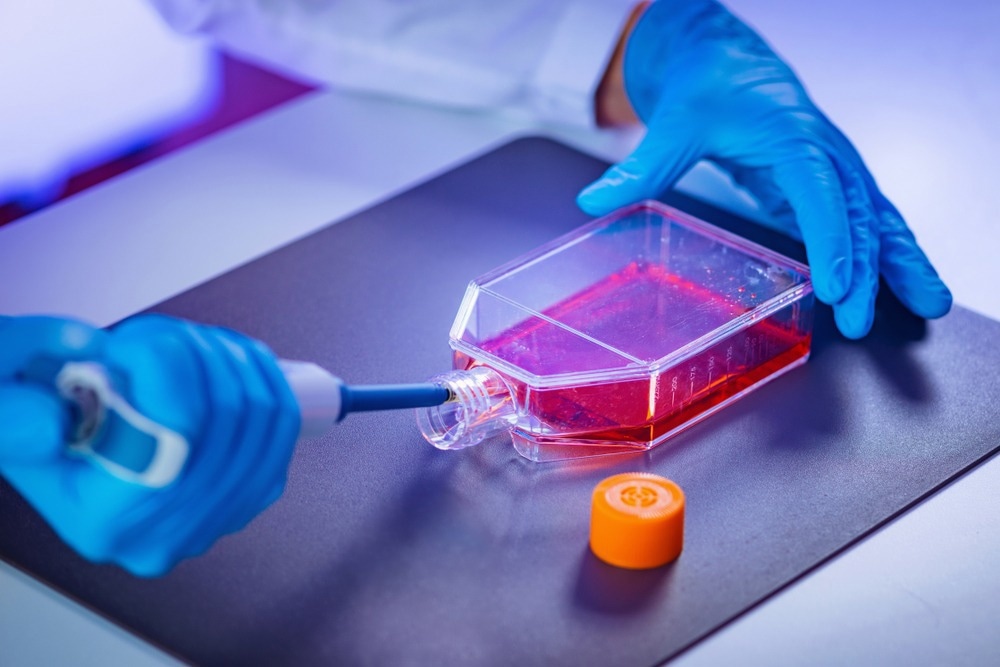
Nanoparticles have many dynamic applications and are synthesized by chemical and biological methods. Biogenic nanoparticles are developed using biological processes using microbial and plant extracts. Recently, biogenic nanoparticles have been applied in tissue engineering (TE) to obtain improved mechanical and biological performances.

Image Credit: Microgen/Shutterstock.com
Synthesis of Biogenic Nanoparticles
Natural sources, such as microorganisms (e.g., bacteria, fungi, yeast, and algae) and plant extracts, have acted as eco-friendly precursors for producing nanoparticles with several potential applications. Both bacteria and fungi are capable of intracellular and extracellular synthesis of nanoparticles.
The biological method is eco-friendly, greener, energy-saving, and a cost-effective approach to nanoparticle synthesis. Another advantage of biogenic nanoparticles is relatively better biocompatibility than other nanoparticles that are synthesized using chemical methods.
Biocompatibility is an essential property of nanoparticles, particularly for biomedical applications. Nanoparticles synthesized using biogenic methods possess many interesting characteristics related to morphology and size. For instance, the size of silver (Ag) nanoparticles synthesized using fungal biomass Verticillium was found to be around 25 nm. These nanoparticles are non-toxic as they allow the fungal biomass to grow continually.
Plant extracts of root, leaf, latex, stem, and seed have been used to synthesize nanoparticles. Notably, plant extracts act as both reducing and stabilizing agents. For instance, the leaf extract of Jasminum sambac was able to synthesize Ag, Au, and Au-Ag alloy nanoparticles. Using this plant extract, triangular, hexagonal, and spherical-shaped Au nanoparticles were synthesized. The bioactive metabolites and enzymes present in the plant extract mediate the synthesis of nanoparticles.
Nanoparticles and Tissue Engineering (TE)
TE involves the growth of new tissues and organs, starting from a base of cells and scaffolds. Scaffolds are three-dimensional (3D) structures that support the cells to grow, proliferate, and differentiate into distinct cell types. TE introduces growth factors into the scaffolds to direct the cells to function desirably. The main goal of TE is to develop fully functional organs or tissues suitable for implantation.
TE techniques face many challenges including the inability of the engineered materials to recapitulate the properties of natural tissues. Several scientific documents have indicated that nanoparticles could alleviate the challenges of TE.
Nanoparticles have been used in TE to obtain superior mechanical and biological performance. The antimicrobial properties of silver nanoparticles and other metallic nanoparticles, electromechanical properties of carbon nanotubes (CNTs), and fluorescence properties of quantum dots have been used in TE. Magnetic nanoparticles (MNPs) have been used to study cell mechanotransduction, gene delivery, and the construction of complex 3D tissues.
The main properties of nanoparticles beneficial in TE are their small size and large surface-to-volume, comparable to small proteins and peptides. These nanoparticles can easily diffuse across membranes and are uptaken by cells. In addition, nanoparticles can mimic the naturally occurring nanosized extracellular matrix (ECM) components of tissues. Besides these properties, biocompatibility and low immunogenicity are the two most important features of nanoparticles for TE applications.
Application of Biogenic Nanoparticles for TE
Nanoparticles are applied in TE for various functions, such as DNA transfection, and increase in the electrical, biological, and mechanical properties of gene delivery and viral transduction. Furthermore, nanoparticles are used to pattern cells to facilitate the growth of various types of tissues for molecular detection and biosensing. Some of the key applications of nanoparticles in TE are discussed below:
Cell Proliferation Rates
Titanium dioxide (TiO2) nanoparticles are used to enhance the cell proliferation rate of cardiac tissue regeneration. Au nanoparticles have exhibited superior biocompatibility and surface modification ability. In TE of bone, Au nanoparticles promote osteogenic differentiation of an osteoblast precursor cell line (MC3T3-E1).
Au nanoparticles also aid in the formation of osteoclast, which is a bone-resorbing cell, from the hematopoietic cell. The size of the biogenic gold nanoparticles, i.e., around 30–50 nm, is extremely important for this function. Implementation of Au nanoparticles and gold nanowires within a scaffold showed a remarkable effect on cell proliferation and synapse formation, essential for an organ transplant. Similarly, incorporating TiO2 nanoparticles in the scaffold associated with human embryonic stem cell-derived cardiomyocytes also revealed increased cell proliferation.
Mechanical and Electrical Properties
Including nanoparticles in nanocomposite polymers, i.e., both in the electrospun fibers and hydrogels, revealed enhanced mechanical properties in TE application, compared to the scaffold without nanoparticles. The addition of TiO2 in a biodegradable patch exhibited a greater tensile strength in reinforcing the scar after myocardial infarction. CNTs are added to nanocomposites due to their extraordinary mechanical properties, tensile strength, and fiber-like structure.
Nanoparticles have been used to enhance the electrical properties of scaffolds, which has proved beneficial in cardiac TE. Adding Au nanoparticles into fibrous decellularized matrices linked to cardiac cells within the scaffolds revealed significant improvement in striation behavior, morphology, and elevated electrical coupling proteins.
Antibacterial Applications
Nanoparticles are also utilized in TE for their antibacterial applications. For instance, the antibacterial properties of poly(3-hydroxybutyrate-co-3-hydroxyvalerate) (PHBV) nanofibrous scaffolds containing Ag were documented. Subsequently, these were effectively used in joint arthroplasty.
Gene Delivery
In TE, gene delivery technology plays a crucial role in targeting matured cells or stem cells. In the case of gene therapy, it is important to develop an appropriate vector system with high specificity to unhealthy cells, low cytotoxicity, and a greater gene transfection efficiency. The efficacy of gene delivery has been substantially improved using self-assembled nanoparticles. DNA transfection using cationic liposomes and viral transduction are two methods used in gene delivery linked to TE application.
Constructing 3D Tissues
Post-organ transplantation, nanomaterials, having unique visual and magnetic properties, are used as suitable agents for monitoring cellular performance in vivo. These are used to develop 3D tissue-engineered scaffolds for bone, skin, vasculature, and other tissues.
Magnetic force-based tissue engineering (Mag-TE) has been developed that uses cells labeled with magnetic nanoparticles (MNPs) to construct tissues. Based on Mag-TE analysis, the B-cell lymphoma 2 (Bcl-2) protein improves the development of synthetic skeletal muscle tissue constructs with an elevated cell density. Mag-TE has also been recently used as a cell sheet for the engineering of liver tissue.
References and Further Reading
Mughal, B. et al. (2021) Biogenic Nanoparticles: Synthesis, Characterisation and Applications. Applied Sciences. 11(6), p. 2598. https://www.mdpi.com/2076-3417/11/6/2598
Patil, S. and Chandrasekaran, R. (2020) Biogenic nanoparticles: a comprehensive perspective in synthesis, characterization, application and its challenges. Journal of Genetic Engineering and Biotechnology, 18, p. 67. https://jgeb.springeropen.com/articles/10.1186/s43141-020-00081-3
Sharma, D. et al. (2019) Biogenic synthesis of nanoparticles: A review. Arabian Journal of Chemistry. 12 (8), pp. 3576-3600, https://www.sciencedirect.com/science/article/pii/S1878535215003147
Hasan, A. et al. (2018) Nanoparticles in tissue engineering: applications, challenges and prospects. International Journal of Nanomedicine. 24(13), pp. 5637-5655. https://www.ncbi.nlm.nih.gov/pmc/articles/PMC6161712/. PMID: 30288038; PMCID: PMC6161712.
Vieira, S. et al. (2017) Nanoparticles for Bone Tissue Engineering. Biotechnology Progress. doi.org/10.1002/btpr.2469
Disclaimer: The views expressed here are those of the author expressed in their private capacity and do not necessarily represent the views of AZoM.com Limited T/A AZoNetwork the owner and operator of this website. This disclaimer forms part of the Terms and conditions of use of this website.