Digital Holographic Microscopy (DHM) powers the Lyncée Transmission series, which uses interferometric resolution for quantitative imaging and measuring of transparent and semi-transparent materials. After holograms are captured and digitally recreated, 3D information is extracted. This information is then analyzed by specialized software to produce original and significant insights for applications in materials science and the life sciences.
DHM gives assessments of morphology and intracellular content (dry mass) and allows label-free imaging in the life sciences. Applications in cell viability, dynamics monitoring, and 4D cell tracking are made possible by these metrics, which are endogenous indicators of biological processes and cell health.
In materials science, DHM gives information on surface topography, thickness variation, and intra-sample features such as defects and refractive index variations. It also monitors dynamic changes and 3D positions over time. It is compatible with the stroboscopic module, which allows for the measurement of optical MEMS, displays, and acoustofluidic devices.
Key Features
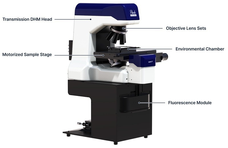
Image Credit: Park Systems
Superior DHM Performance
- Single shot full-field imaging
- Up to 100 kfps acquisition rate
- Millisecond time resolution
Address All Experimental Needs
- Multi-objective, multi-wavelength
- Fully upgradeable
- Several options tailored to specific applications
Lyncée Transmission Series: Workflow
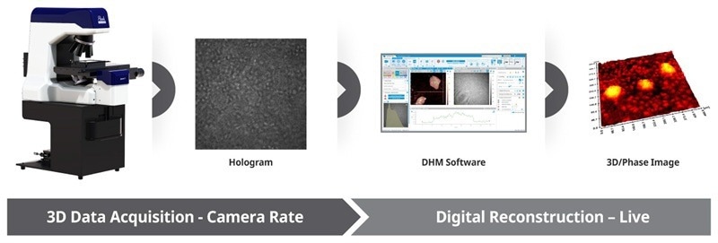
Image Credit: Park Systems
Data acquisition encodes all of the sample's information in the recorded hologram in a single shot at camera rate speed. The captured hologram is then numerically rebuilt in real time. Numerical reconstruction allows users to refocus the phase image after acquisition and obtain quantitative information that provides insights into the monitored sample.
Transmission DHM Models
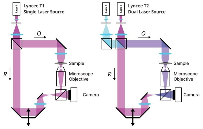
Image Credit: Park Systems
The Lyncée Transmission series (Lyncée T1, T2) includes single- and dual-laser source configurations designed for accurate optical topography. Each model is an excellent tool for its intended application: Lyncée T1 for most applications and Lyncée T2 for large-step-height samples.
Objective Lens Sets
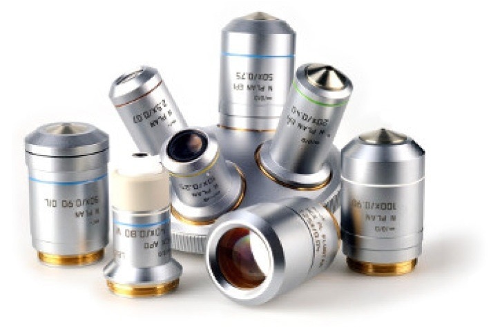
Image Credit: Park Systems
Up to six objectives with magnifications ranging from 1.25 × to 100 × (air and oil immersion) are supported by the objective turret, making it simple to transition between high-magnification examination and wide-field overview.
Motorized Stage
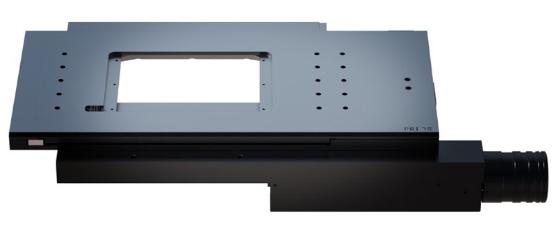
Image Credit: Park Systems
Automated acquisitions at predetermined XYZ locations can record several sites without user involvement, thanks to the optional motorized stage. For large samples, the motorized stage also enables stitching of multiple acquisitions to obtain a broader field of view. It works with conventional multi-well microplates (up to 384 wells) for high-content and high-throughput screening tests in the life sciences.
Fluorescence Module
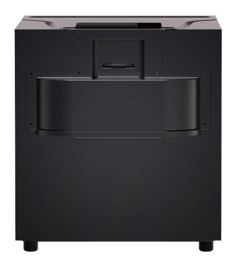
Image Credit: Park Systems
The optional fluorescence module allows for simultaneous quantitative phase imaging and epifluorescence observation using the same objective lens when paired with the Lyncée Transmission series. For uses like cardiotoxicity screening and single-cell analysis, this enables a direct connection between label-free DHM measurements and fluorescence reference assays. Multiple light sources and cameras are supported by the plug-and-play module, and crosstalk-free operation is ensured by dedicated filters.
Environmental Chamber
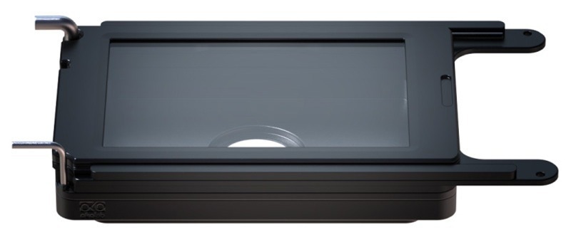
Image Credit: Park Systems
The ideal supplement to non-perturbing multi-day time-lapse experiments. It provides precise control over temperature, O2, CO2, and humidity, comparable to a full incubator. An optional gas mixer allows for O2 control in hypoxia and hyperoxia investigations.
Principle of Digital Holographic Microscopy
DHM Technology
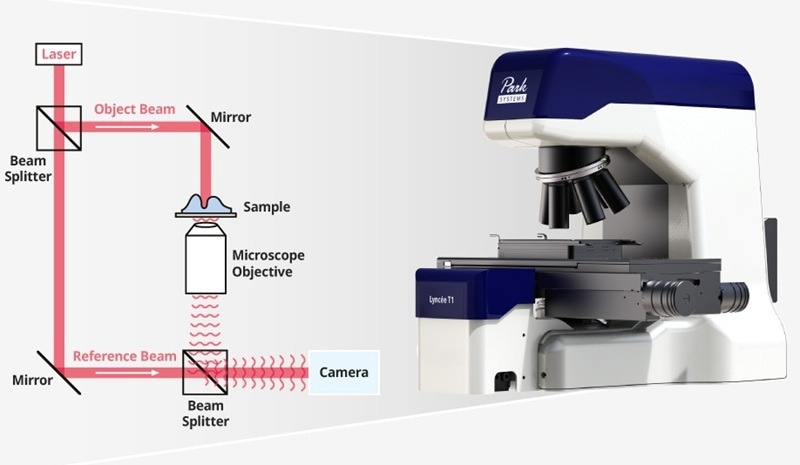
Image Credit: Park Systems
The Lyncée Transmission series offers real-time Quantitative Phase Imaging (QPI) of living cells for use in life sciences applications. Biological characteristics of individual cells, such as viability, confluency, and cell dynamics, are then obtained by segmentation.
Applications
Micro-Optics
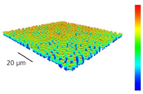
Image Credit: Park Systems
Micro-Fluidics and PIV
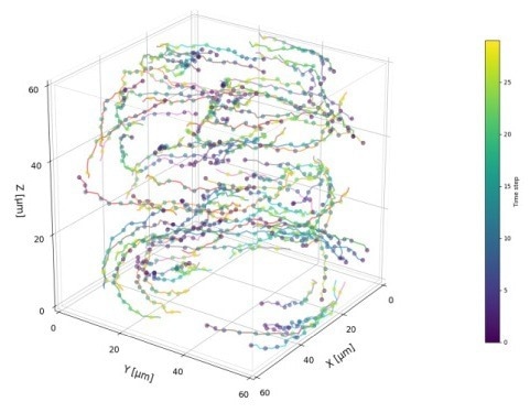
Image Credit: Park Systems
Acoustofluidics
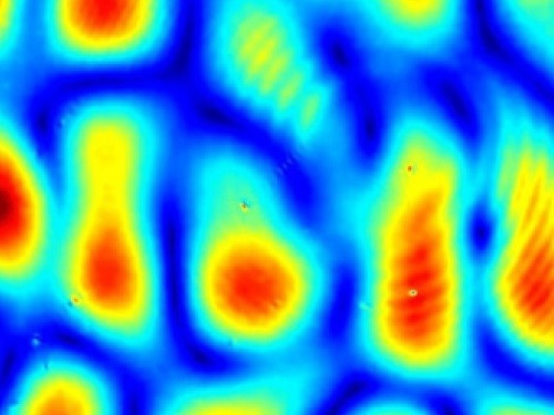
Image Credit: Park Systems
Displays
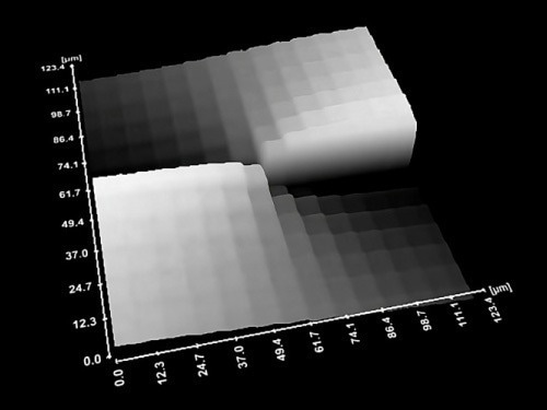
Image Credit: Park Systems
Lipid Droplet Quantification
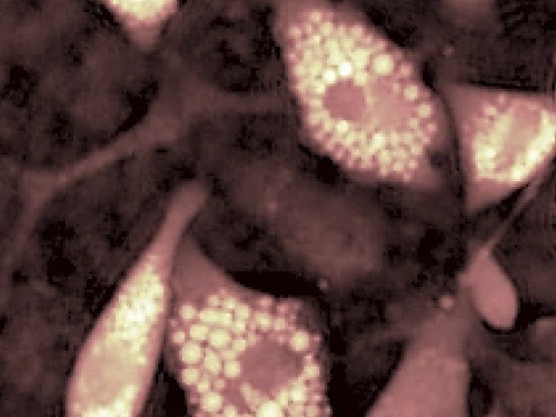
Image Credit: Park Systems
Cell Dynamics
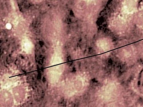
Image Credit: Park Systems
Correlation between DHM and Fluorescence
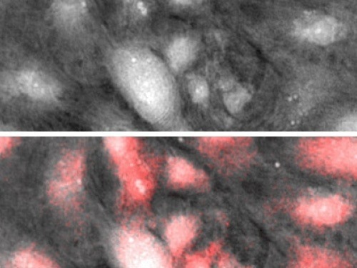
Image Credit: Park Systems
Cell Migration Screening
Image Credit: Park Systems
Softwares
Koala Acquisition and Analysis Software
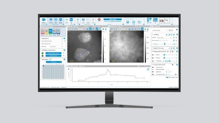
Image Credit: Park Systems
Koala is the primary platform for DHM hardware control, picture capture, processing, and data visualization, with a real-time, user-friendly interface, GPU-accelerated performance, and configurable image and analytical data export options.
Cell Analysis Tool (CAT) Software for Bioanalysis
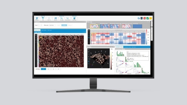
Image Credit: Park Systems
A comprehensive workflow from image acquisition to data visualization and report production is provided by CAT, an analysis program created specifically for life sciences research that integrates seamlessly with our exclusive acquisition software, Koala.