Flow cytometry is a technique used for analyzing and counting microscopic particles such as bacteria, cells and other micro-sized particles.
 Optofludic flourescent imaging cytometry on a mobile phone
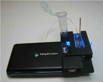
A research team from the BioPhotonics Laboratory at the UCLA Henry Samueli School of Engineering and Applied Science has developed a light-weight and cost-effective optofluidic fluorescent cytometry device to quickly image body fluids for analyzing or counting of cells. This compact optofluidic platform integrates florescent microscopy and imaging cytometry and can be integrated in a mobile phone.
The research team integrated simple optical components to develop the optofluidic platform weighing just 18 g. It consists of simple batteries, one compact lens, one plastic color filter and two LEDs.
The microfluidic device was positioned above a separate lens that is made to contact the present camera module of the mobile phone. This enables the CMOS sensor-chip of the phone to map a complete cross-sectional image of microfluidic device. A sample liquid is constantly directed across a microfluidic channel through a syringe pump.
LEDs based on butt-coupling technology illuminates the device from the sides. The excited light is then directed inside the cross-section of the microfluidic device to excite the the samples in the imaging liquid evenly. The optofluidic pumping system uses the plastic absorbing filter to form a dark background that is required for fluorescent imaging. Additionally, tracking algorithms and video post processing and contour detection are employed for labeling and counting the particles or cells that passes through the device chip. The researchers tested the device to measure the density of WBCs in human blood samples.
This study was sponsored by the Vodafone Americas Foundation, the Gates Foundation, the Office of Naval Research, the National Institutes of Health, and the National Science Foundation.