A collaborative team of researchers from six Italian research agencies has devised a novel microscopy technique that provides a sharp image of the brain’s neural networks.
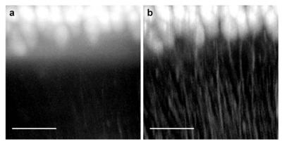 This shows Purkinje cells from a mouse cerebellum imaged (a) with light-sheet microscopy and (b) with the significantly higher contrast provided by confocal light-sheet microscopy (Credit: Optics Express/European Laboratory for Non-Linear Spectroscopy, University of Florence, Italy)
The new technique involves sight correction of an existing imaging technique plagued by blurred images and can be compared to the correction carried out on the Hubble Space Telescope to remedy its blurred vision.
Single plane illumination microscopy (SPIM) or Light-sheet based microscopy (LSM) is an imaging technique for biological samples in which a laser beam focused to just few microns is used to illuminate the sample from the side with a thin sheet of light. A lens is then used to focus the fluorescence radiated off the illuminated sample upward to a digital camera. This technique allows only a portion of the sample to be imaged. A 3D image of the complete sample can be obtained by piecing together 2D images of different sections of the sample.
Researchers have been trying to map the brain’s billion-fold neural network. LSM has proven useful in imaging brain tissues from mice but has given only blurry images for whole brain samples as the samples scatter emitted light resulting in background fluorescence that blurs the image. To address this problem, the Italian researchers integrated LSM with confocal microscopy which involves the use of a filter to eliminate photons straying from the thin sheet’s single plane.
The new technique labeled as confocal light sheet microscopy or CLSM produced sharp images without the need for multiple imaging and data processing techniques. In order to extend the application of this technique to the human brain, it is necessary to overcome the problem of staining fixed tissue fluorescently.