PerkinElmer, Inc., a global leader focused on improving the health and safety of people and the environment, today announced the launch of its Mantra™ Quantitative Pathology Workstation with inForm®Image Analysis Software for cancer immunology and immunotherapy research.
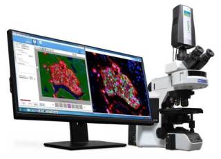 Mantra™ quantitative pathology workstation with inForm® image analysis software enables easy visualization, quantification and phenotyping of multiple types of immune cells simultaneously in intact FFPE tissue sections for cancer immunology research. (Photo: Business Wire)
Unleashing the body’s immune system to fight cancer through promising new cancer immunotherapy methodologies is one of the most promising fields of cancer research. Until now, researchers have only been able to obtain limited information about the types and distribution of immune cells inside and around a tumor. PerkinElmer’s solution enables researchers to investigate the spatial relationships among multiple types of immune cells simultaneously within and around solid tumors. The knowledge resulting from this research may eventually help physicians to better evaluate an individual’s immune response to cancer and could potentially lead to the development of new cancer immunotherapy treatments.
PerkinElmer’s Mantra workstation is an easy-to-use, compact, quantitative pathology imaging solution for quantifying biomarkers and protein expression in situ. It enables the development of multiplexed immune cell and protein expression profiling assays for cellular phenotyping in the tumor and tumor microenvironment of standard formalin-fixed, paraffin-embedded (FFPE) tissue sections.
The Mantra workstation was developed for use with PerkinElmer’s comprehensive cancer immunology research workflow solution. It readily integrates with PerkinElmer’s Opal™ multiplexed immunohistochemistry reagents and inForm software’s new quantitative per-cell analysis for phenotyping immune cells in situ. The system is designed to augment how a pathologist works at the microscope so that users can quickly and easily navigate slides at different magnifications. They can also acquire images with minimal microscope manipulation to visualize the distributions of multiple immune cell phenotypes simultaneously.
“Cancer immunology research provides hope for better outcomes, especially as new drug candidates make their way through the development process,” said Brian Kim, President, Life Sciences & Technology, PerkinElmer. “PerkinElmer is transforming how researchers can see the distributions of immune cells in the tumors they are treating, which can have a significant impact on their understanding of the immune system’s role in cancer.”