A novel microscopy technique called magnetoelectric force microscopy (MeFM) was developed to detect the local cross-coupling between magnetic and electric dipoles. Combined experimental observation and theoretical modeling provide understanding on how a bulk linear magnetoelectric effect can be realized in a new family of materials.
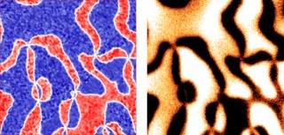 Left: Piezo-response force microscopy image of ferroelectric domains in hexagonal erbium manganite. Image size: 16 x 16 ìm2. Blue (red) color denotes domains with up (down) electric polarization. Right: Magnetoelectric force microscopy image showing a map of electric field induced magnetic response of the same area as that in the left panel. Dark (bright) contrast denotes positive (negative) magnetic response. The one-to-one correspondence between these two images confirms that the sign of electric-induced magnetic response is determined by the polarization state. Image courtesy of Weida Wu
The Impact
The development of local probes to visualize magnetoelectric coupling at mesoscopic scales enables the explorations of emergent phenomena in new materials with multiple coupled orders. The cross-coupling between the magnetic and electric dipoles holds promise for conceptually novel electronic devices for applications such as low-power memory or high sensitivity magnetic sensors.
Summary
The magnetoelectric effect originates from the cross-coupling between the magnetic and electric dipoles in insulating magnets. It holds promise for conceptually novel electronic devices such as electric field controlled magnetic memory and compact magnetic field sensors. However, the existence of domains and defects in these ferroic materials strongly influences their macroscopic responses, which calls for development of local probes of the magnetoelectric effect. Researchers at Rutgers University developed a novel MeFM technique that combines magnetic force microscopy with in situ modulation of high electric fields. This microscopy technique enables direct visualization of the magnetoelectric response of the domains in multiferroic materials (e.g., hexagonal manganites). The interesting observation of the sign change of magnetoelectric response at each structural domain wall was explained by theorists at Cornell and Groningen (Netherlands) using symmetry analysis and phenomenological modeling, which provide compelling evidence that the magnetoelectric coupling is mediated by a periodic lattice distortion. Furthermore, the MeFM results revealed a gigantic enhancement of the magnetoelectric effect when the magnetic order can rotate freely, suggesting a viable way to enhance magnetoelectric couplings for potential multifunctional applications. The detection of magnetoelectric response at mesoscopic scales not only allows direct visualization of magnetoelectric domains, but also opens up explorations of exciting emergent phenomena in multifunctional materials with multiple coupled orders.
Funding
DOE Office of Science, Basic Energy Sciences and the National Science Foundation (Cornell Materials Research Science and Engineering Center).
Publications
Y. Geng, W. Wu, “Magnetoelectric force microscopy based on magnetic force microscopy with modulated electric field.” Review of Scientific Instruments 85, 053901 (2014). [DOI: 10.1063/1.4874006]
Y. Geng, H. Das, A.L. Wysocki, X. Wang, S.W. Cheong, M. Mostovoy, C.J. Fennie, W. Wu, “Direct visualization of magnetoelectric domains.” Nature Materials 13, 163–167 (2014). [DOI: 10.1038/NMAT3813]
H. Das, A.L. Wysocki, Y. Geng, W. Wu, C.J. Fennie, “Bulk magnetoelectricity in the hexagonal manganites and ferrites.” Nature Communications 5, 2998 (2014). [DOI: 10.1038/ncomms3998]