The National Institutes of Health awarded a University of Arkansas biomedical engineer a new $744,992 grant to improve imaging and early detection of chronic wounds and guide treatments.
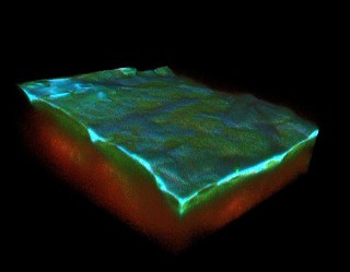 In this three-dimensional reconstruction of skin, acquired through multi-photon microscopy, the outer layer, or epidermis, is identifiable through the natural auto-fluorescence of NADH, FAD and keratin, as seen in blue and green. The dermal layer is detectable in red. Photo by University of Arkansas
Kyle Quinn, assistant professor of biomedical engineering, uses multi-photon microscopy to create 3-D, micro-scale images of chronic wounds to measure different wound properties. The images reveal the metabolism and structure of skin layers and wound regions so that physicians can diagnose chronic wounds and determine appropriate treatment.
Chronic, non-healing wounds are caused by poor circulation, neuropathy, immobility and other factors. They affect millions of Americans and require advanced care with annual costs in billions of dollars. Infection is often a problem, and mortality rates for people with chronic wounds can exceed that of many cancers. Clinicians struggle because there are not optimal methods to diagnose a wound or evaluate appropriate therapeutic interventions.
“It’s currently a wait-and-see approach,” Quinn said. “After an initial diagnosis or checkup, the patient will come back weeks later, and the clinician will evaluate whether the wound has closed or begun closing. If it hasn’t, then the clinician will have to try some other treatment.”
With multi-photon microscopy, Quinn can exploit the intrinsic fluorescence of NADH and FAD, two cellular metabolic cofactors found in chronic wounds. These cofactors are necessary for most metabolic pathways. Multi-photon microscopy allows Quinn to build images of wound sections so he and others can detect and assess metabolic changes.
Data from a pilot phase of the research, funded by an earlier NIH award and accepted for publication in the Journal of Investigative Dermatology, indicated the ability to see differences in the metabolism of diabetic and non-diabetic wounds in frozen tissue samples. With the new grant, Quinn will non-invasively monitor individual wounds in live animals throughout the healing process. By the end of the three-year grant, he hopes to develop a suite of quantitative biomarkers to predict wound chronicity and to guide therapy. The long-term goal is to test the method in a clinical setting.