Oct 26 2016
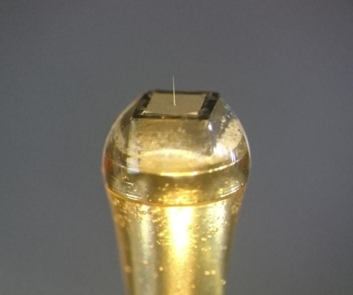
 Extracellular needle-electrode with a diameter of 5µm mounted on a connector. CREDIT: Copyright (C) Toyohashi University Of Technology. All rights reserved.
Researchers from the Department of Electrical and Electronic Information Engineering and the Electronics-Inspired Interdisciplinary Research Institute (EIIRIS) at Toyohashi University of Technology have developed needle-electrodes with a diameter of 5 µm on block modules with the dimensions 1 mm x 1 mm.
The tiny needles could help to solve the mysteries of the brain and can open the door to develop a brain-machine interface. On 25 October 2016, the outcomes of the research were published in Scientific Reports.
As the human brain has highly complex neuron networks, needle-electrode devices made of microfabricated silicon were anticipated to be a breakthrough in enabling the recording and analysis of electrical activities of microscale neuronal circuits that occur in the brain.
However, techniques of developing smaller needles, for example, needles with diameter < 10 µm, can play a vital role in minimizing brain tissue damage. Apart from needle geometry, the size of the device substrate must be reduced to minimize the total tissue damage as well as to improve the accessibility of the electrode in the brain. Therefore, such electrode technologies will achieve new experimental neurophysiological concepts.
The recording made on mouse cerebrum cortices exhibited that the discrete microneedles fabricated on the block modules are minute enough to be used in the narrow spaces of brain tissue. The block module exceptionally enhances the design variability in the packaging, thus providing a number of in vivo recording applications.
Hirohito Sawahata, the first author and Assistant Professor, and Shota Yamagiwa, co-author and researcher, explained “We demonstrated the high design variability in the packaging of our electrode device, and in vivo neuronal recordings were performed by simply placing the device on a mouse’s brain. We were very surprised that high quality signals of a single unit were stably recorded over a long period using the 5µm-diameter needle.”
Our silicon needle technology offers low invasive neuronal recordings and provides novel methodologies for electrophysiology; therefore, it has the potential to enhance experimental neuroscience. We expect the development of applications to solve the mysteries of the brain and the development of brain-machine interfaces.
Takeshi Kawano, Associate Professor, Toyohashi University of Technology