Mar 14 2019
In day-to-day life, blinking lights can be used for sending signals––for instance, that a car is about to turn. At present, tiny “blinkers” that show single molecules of RNA or protein within cells based on the duration and frequency of every flash have been devised by scientists.
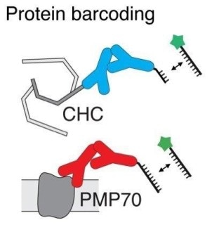 To image two proteins (CHC and PMP70) with the same fluorophore (green star), researchers targeted the proteins with two different lengths (8 or 9 nucleotides) of DNA sequences attached to antibodies (Y-shaped molecules). (Image credit: American Chemical Society)
The study, reported in Nano Letters, an ACS journal, could pave the way for researchers to notice the areas of various biomolecules in a cell at the same time, potentially leading to improved diagnostics and treatments.
Recently, researchers developed superior-resolution microscopes that have the potential to image few-nanometer-sized single molecules. In general, to distinguish between a particular nucleic acid or protein, a fluorescent probe that binds to the molecule and discharges light of specific wavelength is added.
However, due to the possibility of the emission wavelengths of various fluorescent probes to overlap, often, only three or four unique proteins or nucleic acids, rather than the thousands that are present in the cells, could simultaneously be detected by the scientists. Ralf Jungmann and his co-workers opined to use fluorescent probes that could blink with light at variable time and frequency to observe many biomolecules at a time. Hence, this led them to image various molecules using a single fluorophore.
The scientists developed a system based on complementary sequences of DNA that come together to bind a fluorophore with a target biomolecule and then become separated again, producing a blinking fluorescent signal. By changing the length and number of DNA sequences bound to the target, the scientists were able to adjust the duration and frequency of the blink.
The researchers tested their system in the cells by imaging two different RNA molecules and two proteins. Later, they used three fluorescent probes to image 124 unique DNA structures that had many target DNAs in order that they blinked at different frequencies. The researchers reported that the process took only a few minutes and had an accuracy of 97.6%.
This study received funding from the German Research Foundation, the European Research Council, the Allen Distinguished Investigator Program, the Max Planck Society, the Max Planck Foundation, the National Science Foundation, the National Institutes of Health, the Graduate School of Quantitative Biosciences Munich (QBM), and the International Max Planck Research School for Molecular Life Sciences (IMPRS-LS).