Feb 8 2021
In the early 1980s, the advent of scanning probe microscopes revolutionized imaging, paving a way into the nanoscale realm.
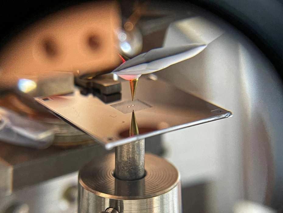
 Photograph of the experimental setup. The separation of the islands is around half a millimeter. Image Credit: David Hälg and Shobhna Misra, ETH Zurich.
The main concept is to scan an extremely sharp tip over a substrate and to record the strength of the interaction between tip and surface at each location. As far as scanning force microscopy is concerned, this interaction is—as the name suggests—the force present between tip and structures on the surface.
Normally, this force is found out by quantifying how the dynamics of a vibrating tip vary as it scans over objects that are deposited on a substrate. A general analogy is tapping a finger across a table to sense the objects placed on the surface.
A research team under the guidance of Alexander Eichler, a Senior Scientist in the group of Professor Christian Degen at the Department of Physics of ETH Zurich, changed this paradigm back to front. In the Physical Review Applied journal, they report the first ever scanning force microscope where the tip is at rest, whereas the substrate with the samples on it vibrates.
Tail Wagging the Dog
Performing force microscopy by 'vibrating the table under the finger' might appear like making the complete procedure a lot more complex. However, mastering the complication of this inverted method pays off well.
The new technique promises to drive the sensitivity of force microscopy to its basic limit, beyond what can be anticipated from additional enhancements of the traditional 'finger tapping' method.
The key to excellent sensitivity is the selection of substrate. The 'table' used in the experiments performed by Eichler, Degen, and their colleagues is a perforated membrane formed of silicon nitride, measuring just 41 nm in thickness. Collaborators of the ETH physicists, the team of Albert Schliesser at the University of Copenhagen in Denmark, have determined that these low- mass membranes are excellent nanomechanical resonators with extreme 'quality factors.'
Most significantly, as soon as the membrane is tipped on, it vibrates millions of times, or more than that, before coming to rest. These unique mechanical properties make it beneficial to vibrate the 'table' instead of the 'finger,' at least in principle.
New Concept Put to Practice
Converting this theoretical potential into experimental capability is the goal of a current project between the teams of Degen and Schliesser, with theoretical support from Dr Ramasubramanian Chitra and Professor Oded Zilberberg of the Institute for Theoretical Physics at ETH Zurich. As a stepping stone on that journey, the experimental teams have currently illustrated that the idea of membrane-based scanning force microscopy functions in a real device.
Especially, they demonstrated that neither loading the membrane with samples nor bringing the tip to within a distance of a few nanometers compromises the superior mechanical properties of the membrane.
But as soon as the tip reaches closer to the sample, the amplitude or frequency of the membrane gets altered. For quantifying these variations, the membrane includes not just an island where tip and sample interact, but also another one—mechanically linked to the first one—from where a laser beam can be reflected partially, to offer a sensitive optical interferometer.
Quantum is the Limit
The researchers put this setup to work and successfully resolved tobacco mosaic viruses and gold nanoparticles. Such images act as a proof of principle for the novel microscopy concept, but they do not yet drive the abilities into new territory. However, the destination is just there.
The team plans to integrate their novel method with a technique called magnetic resonance force microscopy (MRFM), to allow magnetic resonance imaging (MRI) with a resolution of single atoms, thereby offering exclusive understanding, for instance, about viruses.
Atomic-scale MRI would be one more revolution in the field of imaging, thus integrating optimal spatial resolution with highly specific chemical and physical data related to the atoms imaged. To achieve that vision, sensitivity close to the basic limit provided by quantum mechanics is required.
The researchers believe that they can achieve such a 'quantum-limited' force sensor, via additional advances in both measurement methodology and membrane engineering. The evidence that membrane-based scanning force microscopy is viable gets the challenging goal a big step closer.
Journal Reference
Hälg, D., et al. (2021) Membrane-Based Scanning Force Microscopy. Physical Review Applied. doi.org/10.1103/PhysRevApplied.15.L021001.