Platinum (Pt) octahedral mono-rim nanoframes were prepared by edge-selective Pt deposition using gold (Au) nanoparticles as a sacrificial template. The Au adlayers growth on Pt skeletons was based on Frank-van der Merwe mode, forming well-developed and sharp edges.
Selective deposition of Pt on boundaries(inner/outer) can help tune the geometry of Au patterning. Ultimately, selective etching of Au led to Pt octahedral dual rim nanoframes with homogeneous shapes and sizes. Furthermore, coating Au around Pt frames endowed the nanoframes with plasmonic features.
Thus, the fabricated plasmonic dual-rim engraved nanoframes possessed light entrapping capacity through surface-enhanced Raman scattering (SERS) on their surface and served as nanoprobes in biosensing.
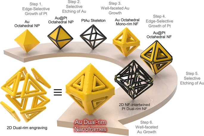
Figure 1. Schematic illustration of the synthesis strategy using multistep chemical reaction conditions. Multiple synthetic pathways for 2D nanoframes (NFs)-engraved octahedral 3D NFs by stepwise chemical toolkits. © Hilal, H. et al. (2022)
Fabrication of Plasmonic Nanoframes
Nanocrystals with complex structures have different physicochemical properties and are applied in nanoscience. Among different types of nanocrystals, nanoframes have found their applications in sensing and imaging due to their high exposed surface and light-matter interactions of their ridges and inner void, promoting the analyte sensing capacity.
The synthesis of nanoframes reported to date was limited to 3D mono-rim nanoframes, which also limited the utilization of their structural functionality. On the other hand, synthesizing nanoframes with complex architecture is challenging.
Furthermore, it is challenging to control the surface structure of nanoframes when their ridges are reduced to a few nanometers of thickness. Only a few previous studies have attempted to resolve specific facets exposed on the ridges of nanoframes. To this end, it is of critical importance to expand the capacity of a synthetic protocol to precisely control the atomic composition and arrangement on the outermost surface of a nanoframe.
Although lithography methods or galvanic replacement reactions were adopted to realize plasmonic nanoframes, the galvanic replacement reaction controlled the nanoframe’s geometrical parameters due to the simultaneous occurrence of reduction and oxidation of the noble metal.
Despite the feasibility of nanostructure production with high precision and fidelity through the top-down lithographical method, this method lacks control over the nanoframe’s physical dimensions in a 3D framework. In addition, fabricating such structures through a bottom-up lithographical method is unfeasible.
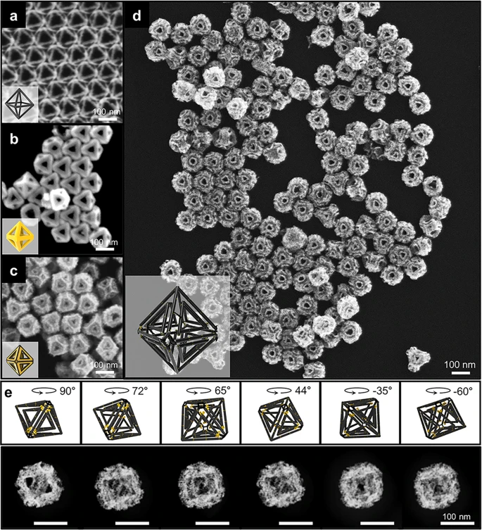
Figure 2. Morphological evolution for octahedral dual-rim NFs using multistep chemical reaction conditions. FE-SEM images of a Pt mono-rim skeletons, b Au mono-rim NFs, c Au@Pt NFs, and d Pt octahedral dual-rim NFs. e Rotational snapshots and cartoons of Pt dual-rim NFs at different angles of −60°, −35°, 44°, 65°, 72°, and 90°. © Hilal, H. et al. (2022)
3D Nanoframes with Dual Rims as Nanoprobes
Previous reports mentioned the controlled synthesis of 2D nanorings with dual rims and an electromagnetic field between outer and inner rims. The synthesis of 3D Au octahedral dual rim nanoframes was also reported with truncated flat terraces on (100) facets.
Thus, the stereoscopic nanoframes with dual rims on every facet could amplify the interaction of particles with light due to gap-induced coupling in the 3D nanostructures. Moreover, the intragap distance would help tune the nanostructure’s light trapping capacity.
The present work demonstrated the synthesis of metallic 3D nanoframes via multi-step wet chemistry. The fabricated nanoframes were composed of a 2D dual rim with a Pt skeleton engraved in a 3D manner. Here, multiple intra-nanogaps of homogeneous shape and size were developed, enhancing the electric near-field confinement.
Previous reports mentioned that the flat terraces facilitated close packing into monolayer arrays contributing to highly sensitive SERS signals. Moreover, an individual particle’s SERS activity depends on its polarization and the orientation of particles, hindering the generation of uniform SERS signals. However, the Au coating on nanoframes in the present work served as a plasmonic component and helped enhance the capacity of near-field focusing due to intra-nanogaps in a single entity, enabling single-particle SERS.
For the first time, the present work reported the construction of frame-frame structures with octahedral nanoparticles in a 3D configuration engraved with 2D dual-rim patterns. Furthermore, the SERS-based biosensing immunoassay demonstrated that the fabricated 3D Au dual-rim nanoframes showed a higher biosensing activity toward human chorionic gonadotropin (HCG) detection than their 2D counterparts, indicating the superior functionality of 3D complex nanoframes.
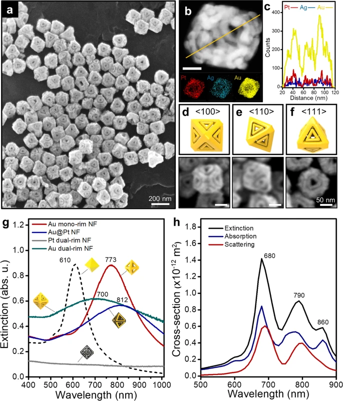
Figure 3. Structural characterization of Au octahedral dual-rim NFs. a A low-resolution FE-SEM image of dual rim-engraved Au NFs. b A HAADF-STEM image of Au dual-rim NFs, energy dispersive spectroscopy (EDS) elemental mapping, and c EDS line mapping for Au, Ag and Pt components. The scale bar denotes 50 nm. d–f FE-SEM images and cartoons viewed from <100>, <110>, and <111> directions, respectively. g Extinction spectra of Au mono-rim NFs, Au@Pt NFs, Pt dual-rim NFs, and Au dual-rim NFs. The numbers denote localized surface plasmon peaks for each particle. h Calculated average optical extinction, absorption, and scattering spectra of Au octahedral dual-rim NFs. © Hilal, H. et al. (2022)
Conclusion
To summarize, a multi-step synthetic method was adopted to fabricate 3D complex nanoframes with dual rims engraved on each facet of octahedral nanoframes. The well-faceted growth was critical to achieving dual rim nanoframes. The layer-by-layer Au growth formed sharp edges, facilitating Pt deposition on the Au mono-rim nanoframes on inner and outer boundaries.
The present synthetic scheme helped achieve complex dual-rim nanoframes with intra-nanogaps between outer and inner rims, enhancing the electric near-field confinement, confirmed through single-particle SERS analysis. Moreover, the 3D nanoframe’s strong near-field focusing capability agreed with the electromagnetic field distribution calculations and surface charge density distribution.
The 3D Au dual-rim nanoframes were applied for SERS-based biosensing immunoassay, proving their potential as nanoprobes in biosensors. Thus, the colloidal chemistry for 3D dual rim nanoframes can contribute toward the fabrication of complex nanoparticles with advanced physicochemical and optical properties.
Reference
Hilal, H. et al. (2022) Three-dimensional nanoframes with dual rims as nanoprobes for biosensing. Nature Communications. https://doi.org/10.1038/s41467-022-32549-w.
Disclaimer: The views expressed here are those of the author expressed in their private capacity and do not necessarily represent the views of AZoM.com Limited T/A AZoNetwork the owner and operator of this website. This disclaimer forms part of the Terms and conditions of use of this website.