This study is led by Prof. Dong Su (Institute of Physics, Chinese Academy of Sciences). Li ion imaging by transmission electron microscopy (TEM) is the “holy grail” in the study of Li ion battery (LIB) materials. Tracking lithiation process in TEM could provide more profound understanding on the electrode degradation mechanism during battery cycling, which accelerates material modification for better performance.
 Research advances in tracking lithiation by analytical TEM, cryo-TEM and in-situ TEM. Image Credit: ©Science China Press
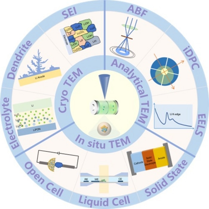
Thanks to recent progress in analytical TEM techniques, such as Annular Bright Field (ABF) method, integrated differential phase contrast and Ptychography, it is now possible to identify Li ion sites inside a crystal. Electron energy loss spectrum could be utilized to detect Li element as well by recognizing its energy loss edge. However, these methods are challenging for beam-sensitive specimens like Solid Electrolyte Interphase (SEI) and Lithium dendrite which are essential to achieve long-term cycling of LIBs. Cryo-TEM has the ability to freeze the specimen to liquid nitrogen temperature, under which condition the high-energy electrons will not damage SEI easily. Moreover, by combining cryo-TEM and tomography method, three-dimensional morphology of Li dendrite could be reconstructed, providing more structural information hidden by two-dimension images.
“Due to the complex kinetics of electrode materials during battery cycling, it is of great importance to observe Li ion diffusion process in real-time. Last decade has witnessed many exciting results through in-situ TEM experiment, including phase transition, ion diffusion, defect evolution and valence change. As conventional dry-cell set-up is used to study the variation of electrode material itself, researchers can now study the reaction between electrode and liquid electrolyte with liquid-cell technique. Combined with emerging methods, we can deeply understand the reaction process and implement the mechanism to the modification of electrode materials.” Su says.
Despite excellent achievements have been made, limitations of these emerging methods still hinder the study of LIB materials. For example, the explanation of ABF and Ptychography images usually requires complex theoretical simulation. Also, continuous observation of electrode materials could damage the specimen during in-situ experiment. Therefore, more efforts should be devoted to improve these methods. Technique combination is one way to overcome the difficulties. For instance, performing low-dose method during in-situ TEM could minimize beam effect. Machine learning is also a path to simplify data analysis procedure and discover the hidden property-structure correlation.