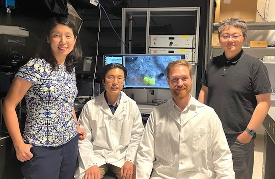
Lan Luan (from left), Robin Kim, Roy Lycke, and Chong Xie are part of a team of researchers with the Rice Neuroengineering Initiative that developed a highly biocompatible, flexible nanoelectrode that can provide intracortical stimulation with a high degree of spatiotemporal stimulus control. Image Credit: Rice University
In rodents, the tiny implantable devices generated stable, long-lasting, and seamless tissue-electrode interfaces with no scarring or degradation, according to research reported in Cell Reports. The devices delivered electrical pulses that more closely matched neuronal signaling patterns and amplitudes than standard intracortical electrode stimulation.
Because of the devices’ great biocompatibility and fine spatiotemporal stimulus control, new brain stimulation therapies, such as neural prostheses for patients with compromised sensory or motor functions, could be developed.
This paper uses imaging, behavioral and histological techniques to show how these tissue-integrated electrodes improve the efficacy of stimulation. Our electrode delivers tiny electrical pulses to excite neural activity in a very controllable manner. We were able to reduce the current necessary to elicit neuronal activation by more than an order of magnitude. Pulses can be as subtle as a couple hundred microseconds in duration and one or two microamps in amplitude.
Lan Luan, Study Corresponding Author and Assistant Professor, Electrical and Computer Engineering, Rice University
The Rice Neuroengineering Initiative investigators’ new electrode design shows a significant improvement over traditional implantable electrodes used to treat conditions like Parkinson’s disease, epilepsy, and obsessive-compulsive disorder, which can cause negative tissue responses and unplanned changes in neural activity.
Conventional electrodes are very invasive. They recruit thousands or even millions of neurons at a time. Each of those neurons is supposed to have their own tune and coordinate in a specific pattern. But when you shock them all at the same time, you’re basically disrupting their function. In some cases that works fine for you and has the desired therapeutic effect. But if, for example, you want to encode sensory information, you need much greater control over the stimuli.
Chong Xie, Study Corresponding Author and Associate Professor, Electrical and Computer Engineering, Rice University
Xie compared standard electrode stimulation to the disruptive effect of “blowing an airhorn in everyone’s ear or having a loudspeaker blaring” in a roomful of people.
“We used to have this very big loudspeaker, and now everyone has an earpiece,” he said.
The capacity to change the frequency, duration, and intensity of the signals could lead to the creation of innovative sensory prosthetic devices.
Neuron activation is more diffuse if you use a larger current. We were able to reduce the current and showed that we have a much more focused activation. This can translate to higher-resolution stimulation devices.
Lan Luan, Study Corresponding Author and Assistant Professor, Electrical and Computer Engineering, Rice University
Luan and Xie are core players of the Rice Neuroengineering Initiative, and their laboratories are also partnering on the advancement of an implantable visual prosthetic device for blind patients.
“Envision one day being able to implant electrode arrays to restore impaired sensory function: The more focused and deliberate is the activation of the neurons, the more precise the sensation you’re generating,” Luan notes.
A previous iteration of the devices has been used to record brain activity.
“We have had a series of publications showing this intimate tissue integration enabled by our electrode’s ultraflexible design really improves our ability to record brain activity for longer durations and with better signal-to-noise ratios,” states Luan, who has been promoted to Associate Professor effective July 1.
The study’s lead authors are electrical and computer engineering Postdoctoral Associate Roy Lycke and Graduate Student Robin Kim.
The research was supported by the National Institute of Neurological Disorders and Stroke (R01NS109361, U01 NS115588) and Rice internal funds.
Journal Reference
Lycke, R., et al. (2023). Low-threshold, high-resolution, chronically stable intracortical microstimulation by ultraflexible electrodes. Cell Reports. doi.org/10.1016/j.celrep.2023.112554.