Schematic diagram of light-induced assembly of extracellular vesicles (EV). Using laser irradiation, the researchers managed to directly detect nanoscale EVs in a cell supernatant within minutes. Image Credit: Takuya Iida, Osaka Metropolitan University
Nanoscale extracellular vesicles (EVs), including exosomes measuring 50–150 nm in diameter, serve crucial functions in intercellular communication. They have also gained recognition as biomarkers for various diseases and as carriers for drug delivery. Hence, the swift and sensitive detection of nanoscale EVs from minute samples is of utmost significance for the early diagnosis of challenging conditions like cancer and Alzheimer's disease.
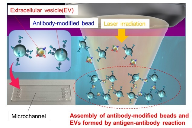
Traditionally, isolating nanoscale EVs from cell culture media involved a complex and time-consuming procedure that included ultracentrifugation.
A research team headed by Director Professor Takuya Iida, Deputy Director Associate Professor Shiho Tokonami, and Assistant Director Professor Ikuhiko Nakase at the Research Institute for Light-induced Acceleration System (RILACS) at Osaka Metropolitan University harnessed the power of laser light to expedite the reaction between nanoscale EVs derived from cancer cells and microparticles modified with antibodies.
They subsequently examined the three-dimensional structure of the resulting aggregates using confocal microscopy. Consequently, the researchers successfully demonstrated the capability to detect approximately 103–104 nanoscale EVs present in a 500 nL sample within a mere 5-minute timeframe.
This research achievement provides a method for ultrafast and ultrasensitive quantitative measurement of biological nanoparticles, offering a foundation for innovative analysis of cell-to-cell communication and early diagnosis of various diseases in the future.
Takuya Iida, Director and Professor, Research Institute for Light-induced Acceleration System, Osaka Metropolitan University
This study received support from various sources, including the JST-Mirai Program (No. JPMJMI18GA, No. JPMJMI21G1), Grant-in-Aid for Scientific Research (A) (No. JP17H00856, No. JP21H04964), JST FOREST Program (No. JPMJFR201O), Grant-in-Aid for Scientific Research (B) (No. JP18H03522), Scientific Research on Innovative Areas (No. JP16H06507), and Grant-in-Aid for Early-Career Scientists (No. JP20K15196) from the Japan Society for the Promotion of Science KAKENHI. Additionally, the Key Project Grant Program of Osaka Prefecture University also provided support for this research.
Journal Reference
Fujiwara, K., et al. (2023). Ultrafast sensitivity-controlled and specific detection of extracellular vesicles using optical force with antibody-modified microparticles in a microflow system. Nanoscale Horizons. doi.org/10.1039/D2NH00576J.