A team of researchers in a joint venture between the University of Illinois and the University of Munich has conducted two studies that explain the chemical and mechanical communications that enable the ribosome, which builds the cell's protein, to integrate a growing protein inside the cellular membrane.
The team in Munich utilized cryo-electron microscopy to capture one precise moment in the procedure, in order to get an understanding of how the ribosome, membrane, membrane channel and new protein bind together. Each structure had been analyzed separately. Co-author Roland Beckmann at the University of Munich said that the Illinois team analyzed the cryo-EM rebuilding at an atomic level, and used simulation to test it. The collaboration combined experimental and computational techniques.
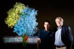 University of Illinois biophysics professor Klaus Schulten (right) and postdoctoral researcher James Gumbart
In order to capture an image of the ribosome's reaction with the membrane, Beckmann's group used nano-disks of membrane bound together with strips of constructed lipoproteins. Stephen Sligar, biochemistry professor at the University of Illinois, designed the nanodiscs. The Illinois group used the cryo-EM pictures and structural data to create an atom-by-atom prototype of the system and insert the proteins into the images provided by the electron microscope, according to biophysics professor Klaus Schulten, who led the Illinois analysis with postdoctoral student James Gumbart.
Some areas of the membrane channel enter the ribosome exit point to direct the protein into the channel, which will string it through the membrane either to remove it or open a side door to steer the protein into the membrane. The images showed that the ribosome seemed to communicate directly with the membrane surface.
At the National Academy of Sciences, Schulten, Gumbart and student Christophe Chipot showed that proteins enter the membrane in two stages. The ribosome first forces the growing protein out into the channel, and then the protein enters the membrane. The chemical energy that the ribosome harvests from other molecules in the cell pushes the cell and enables even highly charged proteins to enter the greasy, nonpolar membrane.
Sopurce: http://www.uillinois.edu