The development of stimulated emission depletion (STED) confocal microscopes has paved the way to overcome the diffraction limit of optical microscopes, which causes a restriction in studying objects having size below 200 nm.
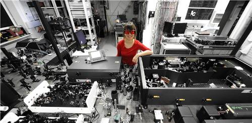 Joanna Oracz (FUW) in her optical lab. (Source: Grzegorz Krzy¿ewski/FUW)
As part of her MA thesis, Joanna Oracz has developed a prototype STED confocal microscope at the Faculty of Physics located at the University of Warsaw. This prototype will be utilized for optical research as well as for biological sample analysis by 2012.
A typical fluorescence confocal microscopy uses a laser beam for scanning a biological sample and agitating dye molecules locally following its introduction into the sample. The light emitted by the agitated molecules is navigated via a filter and tracked by a detector placed after a confocal aperture. The aperture size helps in improving the image contrast by eliminating light from out-of-focus planes.
A STED microscope utilizes one more laser beam called the depletion beam, which triggers stimulated emission from the dye molecules that are illuminated by the beam due to its wavelength. Molecules cannot fluoresce now as their stimulated emission consumes their energy, which in turn avoids other light to pass via the filter and thus they will not be viewed on the captured image. The donut shape of the depletion beam is the basis of the STED method. When the donut-shaped beam and the illuminating beam are matched in space and time, the resulting fluorescence will take place in the sample area situated at the center part of the depletion beam.
Ensuring the overlapping of the two laser beams was the major problem during the development of the prototype. The resolution of the prototype microscope is nearly 100 nm, a value two folds better than that of a typical confocal microscope. Further research is going on for achieving a resolution value of around 60 nm, which makes the prototype to analyze miniscule objects such as dendritic spines of neurons.