Mar 4 2020
A global team of engineers and researchers have made a breakthrough discovery that could additionally augment the application of ultra-thin zeolite nanosheets. These nanosheets are utilized as specialized molecular filters.
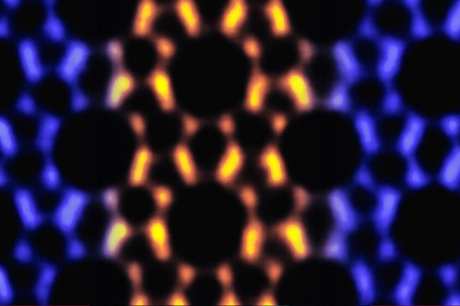
 This image shows atomic-scale details from transmission electron microscopy that reveals the porous structure of an MFI nanosheet, with MEL intergrown in it. Image Credit: Kumar et al., University of Minnesota.
This latest finding can potentially lead to the efficient production of biofuels, plastics, and gasoline. The team was headed by Associate Professor K. Andre Mkhoyan from the University of Minnesota and Professor Emeritus Michael Tsapatsis, now a Bloomberg Distinguished Professor at Johns Hopkins University.
The researchers made this revolutionary discovery of one-dimensional (1D) defects in a two-dimensional (2D) structure of porous material by using a robust, high-resolution transmission electron microscopy (TEM) at the University of Minnesota Twin Cities campus. The porous material used here was a form of zeolite known as MFI.
When the researchers imaged the atomic structure of the MFI nanosheets at an extraordinary detail, they discovered that these 1D defects led to a unique reinforced nanosheet structure that considerably altered the nanosheet’s filtration properties. The study results were published in a peer-reviewed scientific journal, Nature Materials.
TEM imaging of thin zeolite crystal at the atomic-scale has been a long-standing challenge as these crystals are readily damaged under the high-energy electrons, which are needed for atomic-scale imaging.
K. Andre Mkhoyan, Associate Professor, University of Minnesota
Mkhoyan is an expert in sophisticated TEM and the Ray D. and Mary T. Johnson/Mayon Plastics Chair in the Department of Chemical Engineering and Materials Science at the College of Science and Engineering in the University of Minnesota.
He continued, “It requires a deep understanding of the mechanisms of beam damage for zeolite crystals and the doses of electron beam that the zeolite can take. This work pushed the limits of our electron microscopes, where we can reliably produce atomic-resolution images of such extremely thin (just 3-nanometers-thick) zeolite nanosheets with identifiable one-dimensional intergrowths.”
For Prashant Kumar, a graduate in the University of Minnesota Twin Cities College of Science and Engineering, it took almost five years of analysis to detect the slight variations between both materials.
I have been fascinated by the beautiful symmetrical patterns in MFI crystal throughout my Ph.D. work. After staring at noisy images in the TEM for countless hours, I finally saw the symmetry breaking in the TEM images of MFI nanosheets—I knew this was unusual.
Prashant Kumar, Study Lead Author, University of Minnesota
In spite of the slight variations, this knitting of lines of one zeolite inside another one has marked consequences in the potential of ultra-thin nanosheets to detect and selectively convey molecules. This allows for selective separations and catalysis.
Traian Dumitrica (mechanical engineering) and Ilja Siepmann (chemistry), both Professors at the University of Minnesota, performed the simulations to test this performance and pattern. The duo’s findings showed that the knitted materials were less reactive to stress but more selective when it came to isolating molecules based on shape and size.
Membranes produced from these improved nanosheets for the laboratory simulations were created by a team of researchers headed by Tsapatsis. These membranes were tested under industrial conditions and also by Benjamin McCool, head of separations and process chemistry at ExxonMobil.
The latter resulted in the most optimal filtration performance; o-xylene and p-xylene were isolated at five times higher efficiency than reported by the Tsapatsis’ team so far.
MFI zeolite can be defined as a porous structure of oxygen and silicon atoms. Earlier, it was believed to grow with 1D structures, or a zeolite known as MEL, in bulk form. But these defects have never been particularly intergrown or created in 2D nanosheets.
Making ultra-selective thin-film membranes and hierarchical catalysts by fine tuning the frequency and distribution of intergrowths of porous frameworks is a concept introduced by our research group a decade ago.
Michael Tsapatsis, Professor Emeritus and Bloomberg Distinguished Professor, Johns Hopkins University
Tsapatsis continued, “The discovery by TEM of one-dimensional intergrowths in two-dimensional nanosheets and the practical implications suggested by modeling bring the potential of this concept to a new level and suggest new opportunities for targeted synthesis that we have not imagined possible.”
Tsapatsis’ team are now hoping to develop heterostructures of MFI-MEL nanosheets that would not only increase the MEL content but would also push the thin-films’ filtration performance to even greater efficiency, as expected by the laboratory simulations.
For Mkhoyan, who manages the analytical electron microscopy laboratory on the University of Minnesota, where atomic-scale research is routinely performed, the latest discovery presents an opportunity to additionally enhance the way microscopes are used for analyzing nanomaterials down to the atomic-level detail.
The research team members include Ph.D. students and post-doctoral fellows Prashant Kumar, Dae Woo Kim, Neel Rangnekar, Supriya Ghosh, Han Zhang, Meera Shete, and Qiang Xiao.
Faculty members included K. Andre Mkhoyan (from the Department of Chemical Engineering and Materials Science), Evgenii Fetisov and Professor Ilja Siepmann (from the Department of Chemistry); and Hao Xu and Traian Dumitrica (from the Department of Mechanical Engineering); senior research associate Benjamin McCool (from ExxonMobil); and professor emeritus Michael Tsapatsis (from Johns Hopkins).
The study was mainly financed by the National Science Foundation, and the U.S. Department of Energy, while various other partners of the University of Minnesota extended partial support for specific computations and characterizations.
The study is titled “One-dimensional intergrowths in two-dimensional zeolite nanosheets and their effect on ultra-selective transport.”
Atomic-scale imaging reveals secret to thin film strength
This video by University of Minnesota researchers shows one-dimensional intergrowths in two-dimensional zeolite nanosheets and their effect on ultra-selective transport. This breakthrough discovery pushes the limits of microscopy to improve efficiency of fuel and plastic production. Video Credit: University of Minnesota.