Advancements in the early detection of disease has been a field of research that has gathered attention with revolutionary suggestions to further develop medicine.
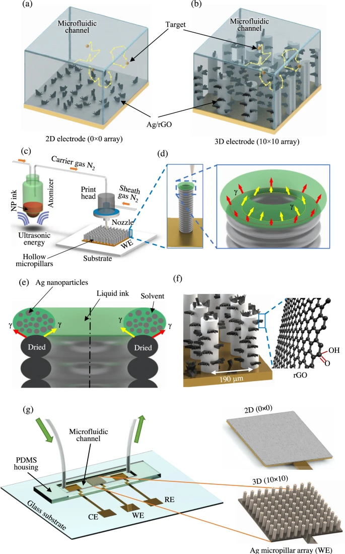
Aerosol Jet nanoparticle 3D printing process for micropillar array electrodes used for dopamine sensing. a, b Schematic representations of Brownian-motion of target molecules for a 2D electrode (0 × 0 array) and a 3D electrode (10 × 10 array) within the microfluidic chamber. c AJ 3D printer with an ultrasonic atomizer and a print head. The metal nanoparticle ink is converted into aerosol of ink droplets in the atomizer. It is then carried to the nozzle via a tube using a carrier gas (N2). A sheath gas, also N2, focuses the aerosol on the substrate at a length scale of 10 μm. d The mechanism of hollow-micropillar formation with concentric toroid-shaped rings of metal nanoparticle (NP) ink printed layer-by-layer. The red and yellow arrows show the surface tension of the liquid ring which helps in the buildup of the structure. Once one layer is printed, it loses solvents due to the heat from the platen and provides a solid base for the next layer of toroid-shaped ring of the ink to be printed. e Cross-sectional view of the printing process shown in (d) where surface tension, γ, provides the fluid dynamic stability required to keep the ring in a stable state. The top liquid ring contains silver nanoparticles with solvents and binders before losing the solvents due to the heat from the platen. The solidified structure is then sintered in an oven as discussed in the “Methods” section. f A schematic showing the silver micropillars with rGO flakes attached to their surfaces. g Schematic of the dopamine sensing device including the PDMS housing, a microfluidic channel, a 3D printed micropillar array (from (c)), tubes for injection and removal of dopamine solution (RE-reference electrode, CE-counter electrode, WE-working electrode). Schematic of a 2D silver sensor fabricated by AJ printing method and used to assess a comparative performance is also shown. © Ali, M., et al (2021)
The Need for Early Disease Detection
Early detection is the main way to prevent and control the spread of disease. As a result, this has become a key research area to ensure early diagnoses can be managed and treated effectively.
However, early disease detection would require the detection of biomarkers with low concentrations, illustrating the need to enhance current sensing techniques, which can be used for a broad range of biomarkers at ultralow or femtomolar concentrations.
The use of these enhanced sensing techniques could also prove to be useful in events such as the SARS-CoV-2 (COVID-19) pandemic, where detecting the coronavirus at ultralow concentrations would be beneficial in stopping the transmissibility of this infectious virus.
Additionally, detection and diagnosis of early biomarkers which can lead to diseases and disorders can prevent any further deterioration of health. This can also be utilized to improve cancer prognoses and ensure patient care is prioritized through personalized medicine.
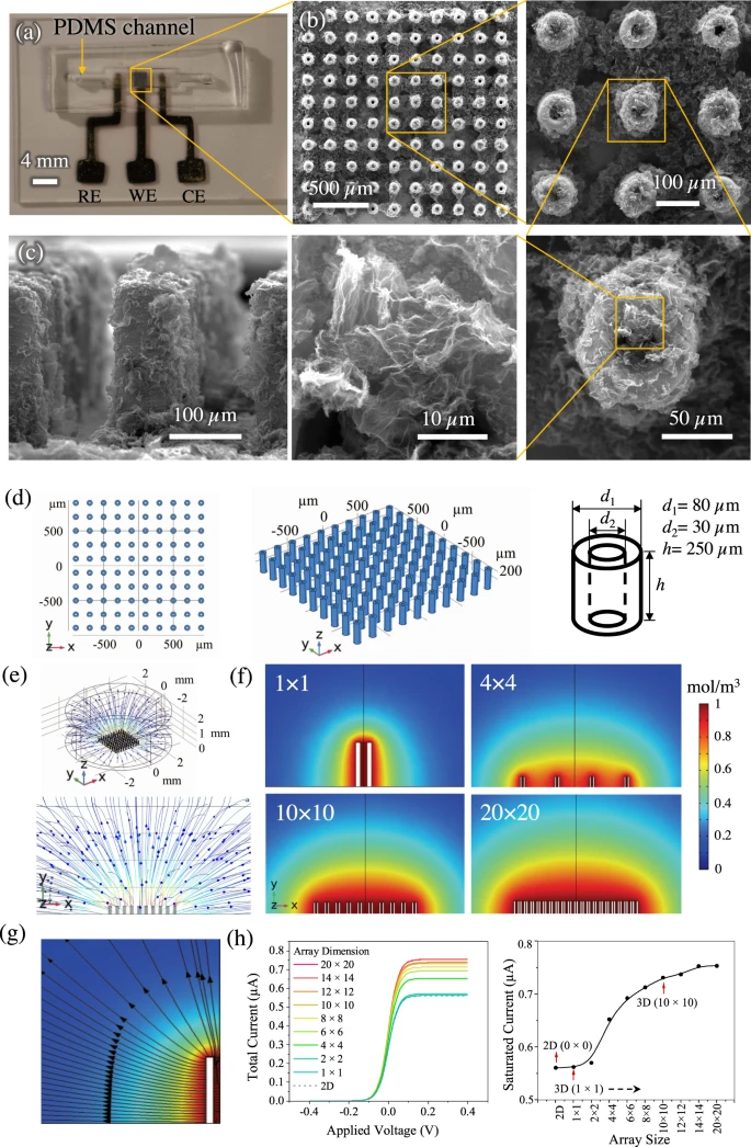
The nano-to-meso multi-length-scale AJ printed biosensing platform. a Optical image of the microfluidic sensing platform for dopamine detection consisting of the electrodes and the PDMS housing. b SEM images of the AJ printed 3D Ag/rGO micropillar array electrode at different magnifications. The top-down view of the micropillars with inner diameter of 28 μm and outer diameter of 80 μm is shown in the micrographs at an increasing magnification. The rGO flakes attached to the silver micropillar surface are also observed. c The side-view of the Ag/rGO micropillar array showing the rGO coating and dimension of the pillar. Two repeated measurements of Fig. 2b, c were performed on two separate sensors. d–h COMSOL simulation of the electrochemical cell. d Model geometry with top-view, 3D perspective view, and dimensions for 10 × 10 configuration. e Characteristic diffusion profiles for oxidation of redox species for 10 × 10 micropillar array configuration for the electrode. f Concentration profiles of diffusing species at the central plane for different arrays of the electrodes—1 × 1, 4 × 4, 10 × 10 and 20 × 20. g Mass flux streamlines for half of a single hollow micropillar (cross-section) which shows the contribution of planar and radial diffusion of electro-species. h One-dimensional (1D) plots of total current obtained from different array configurations varying from 1 × 1 to 20 × 20 as a function of applied voltage (V). The saturation current is obtained for each array configuration. Data for 2D configuration (0 × 0 array) is also included. The data show that for an array with configuration denser than 10 × 10 in the microfluidic chamber, the saturation current does not increase further. The electrode configuration of 10 × 10 array was chosen for this study. © Ali, M., et al (2021)
Increasing Sensitivity to Biomolecules
Increasing the sensitivity to detect biomolecules can be a difficult task; however, electrochemical transduction has been a well-established method for increasing the sensitivity of surfaces for this purpose.
Nanostructuring surfaces to increase sensitivity is also deemed to be an approach with effective results due to the increase in surface area leading to an increase in the reaction sensitivity.
Exploring this avenue has consisted of investigating various nanostructured surfaces, such as DNA nanostructures, bismuth sulfide nanowire microfibers and a range of nanocomposite and nanoparticle combinations with reactive elements.
However, while these methods have advanced detection sensitivity, there are limitations that make these approaches challenging for analyte molecules to make contact with them in the relevant time scale. This is due to the two-dimensional nature of the surfaces that nanostructures are conventionally grown. When the concentration of a typical analyte is low, such as below the picomolar level, the probability of it interacting and making contact with the nanostructures is also decreased.
However, to overcome this challenging obstacle, antibodies which are functionalized with magnetic particles can be used and so when under a magnetic field, they are more likely to be directed into sorting wells, even at low concentrations.
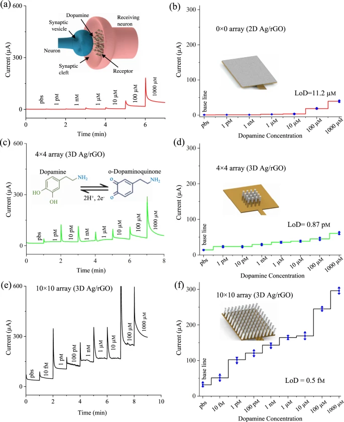
Sensing of dopamine molecules. a Chronoamperometric sensing curves for 2D Ag/rGO (0 × 0 array) sensor. The dopamine (dopamine hydrochloride) concentration is diluted from 1 pM to 1000 μM in pbs solution (pH 7.4) containing 1 mM concentration of ferro/ferricyanide. The measurements were conducted without any enzyme at a fixed sensing potential of −0.1 V for 1 min. Inset of (a) shows a schematic of dopamine molecules released from pre-synaptic neuron to target post-synaptic neuron. b Saturation currents of 2D Ag/rGO sensor plotted against varying dopamine concentrations. c, e Chronoamperometric sensing of multi-length-scale 3D Ag/rGO (4 × 4 array) and 3D Ag/rGO (10 × 10 array) sensors, respectively, at the same condition as that performed for 2D Ag/rGO sensor. A low concentration (10 fM) of dopamine was tested in addition to 1 pM to 1000 μM for 3D Ag/rGO (10 × 10 array) sensor. Inset of (c) shows the biochemical oxidation/reduction reaction of dopamine. d, f Current responses with varying dopamine concentration for multi-scale 3D Ag/rGO (4 × 4 array) and 3D Ag/rGO (10 × 10 array) configurations, respectively. The detection time for all the results in this figure are within 60 seconds. Error bars in Fig. 4b, d, f are the standard deviation of three repeated measurements (n = 3 biologically independent experiments). Error bars, mean ± SD. © Ali, M., et al (2021)
Innovative Research to Overcome Challenges
To mitigate these challenges, innovative researchers have explored using a multi-length scale electrode to enable surfaces of micro- and mesoscale structures to have active interactions with analyte molecules. This type of architecture can be created through 3D printing or additive manufacturing.
The researchers used Aerosol Jet (AJ) nanoparticle printing to fabricate a 3D complex device with multiple hierarchical length-scales, such as within micro to mesoscale.
The benefit of using 3D printing includes the ability to integrate various materials to create complex architecture. This can allow atomically thin nanomaterials such as graphene flakes to be integrated with electrodes resulting in micropatterning as well as ultra-high surface area. The aim of this research is to overcome the limit of detection and achieve the femtomolar level of detecting significant biomolecules.
A significant application for this research includes its use in analyzing a sample such as through hollow silver pillars that protrude into the microfluidic channel containing the analyte.
This critical research can be utilized for the detection of dopamine, which, if detected at low concentrations, can have significant benefits within medicine and disease prevention.
Motor Neuron Diseases
Dopamine is an integral electrochemical messenger found in the brain and has a significant role in controlling motor function and coordination, emotions, cognition, and reward-based behavior. Its critical function can be seen in a more apparent manner when dopamine is dysfunctional, which can lead to a wide range of motor neuron disorders.
When dopamine levels are low, it can cause vasodilation outside of the brain and subsequently result in a disorder such as ADHD (attention deficit hyperactivity disorder), dementia, and Huntington’s. However, dysfunctional high levels of dopamine can cause other diseases such as neuroblastoma, a type of brain cancer.
Current research allows dopamine to be detected at nanomolar concentration levels with an electrochemically grown reduced graphene oxide (rGO)-based electrode detecting dopamine at 0.1 µM through a direct catalytic oxidation reaction.
However, similar to the other detection methods, these involve nanomaterials on 2D surfaces, so researchers in this novel study have utilized graphene to achieve an enhanced level of detection, which would also be able to detect dopamine other analytes.
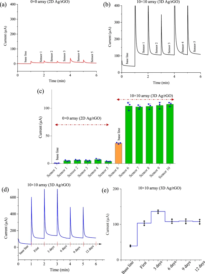
Reproducibility and stability tests of the multi-length-scale sensing platform. Five different sensors for each configuration (2D Ag/rGO and 3D Ag/rGO) were fabricated using the process developed in this work and tested with the same parameters in pbs solution with 1 mM of ferro/ferricyanide for 2D Ag/rGO electrode (a) and 3D Ag/rGO micropillar array electrode (b). The dopamine concentration was set to 20 µM for 2D Ag/rGO sensors and 1 pM for 3D Ag/rGO sensors. c The plot between chronoamperometric currents and the number of sensors for both 2D Ag/rGO and 3D Ag/rGO sensors. d–e The stability test of 3D Ag/rGO sensor was conducted at 1 pM concentration of dopamine by varying time in days while the sensor was kept at 4 °C. Error bars in (c) and (e) are the SD of measurements over at least n = 3 biologically independent experiments. Error bars, mean ± SD. © Ali, M., et al (2021)
Future Therapies
The advancement in detection sensitivity provided by this research has developed the use of nanomaterials to enhance detection sensitivity.
This research that utilizes 3D printing may revolutionize medicine with early detection abilities that can be used to identify biomarkers of disease such as neurodegenerative disorders at an earlier stage to manage and treat them effectively. This can prevent disease progression before critical neurons are damaged, which naturally occurs with time.
The field of neurodegenerative disorders that have not yet been successful in finding effective cures has been provided with a potentially life-changing detection method, aiming to prevent patients from being burdened with very challenging and deadly diseases.
Continue reading: COVID-19 Detection with Carbon Nanotube Based Biosensors
Reference
Ali, M., Hu, C., Yuan, B., Jahan, S., Saleh, M., Guo, Z., Gellman, A. and Panat, R., (2021) Breaking the barrier to biomolecule limit-of-detection via 3D printed multi-length-scale graphene-coated electrodes. Nature Communications, 12(1). Available at: https://www.nature.com/articles/s41467-021-27361-x
Further Reading
Schrötter, A., Magraoui, F., Gröttrup, B., Wiltfang, J., Heinsen, H., Marcus, K., Meyer, H., Grinberg, L. and Park, Y., (2013) Early Diagnosis of Neurodegenerative Diseases - The Long Awaited Holy Grail and Bottleneck of Modern Brain Research - 19th HUPO BPP Workshop. PROTEOMICS, 13(20), pp.2938-2941. Available at: https://analyticalsciencejournals.onlinelibrary.wiley.com/doi/10.1002/pmic.201370164
Disclaimer: The views expressed here are those of the author expressed in their private capacity and do not necessarily represent the views of AZoM.com Limited T/A AZoNetwork the owner and operator of this website. This disclaimer forms part of the Terms and conditions of use of this website.