The prepared nanofibers were composed of poly (ɛ-caprolactone) (PCL) and polylactic acid (PLA) in a 1:1 ratio, the efficiency of which was validated by its drug-loading capacity, morphology, and in vitro drug release.
The ex vivo permeability of the nanofibers was evaluated using chicken pouch mucosa and was subsequently tested for histopathology. Ex vivo permeation highlighted the improved sustained release of the drug and a significant decrease in drug leakage from nanofibers.
Furthermore, scanning electron microscopy (SEM) images revealed the morphology of the prepared nanofibers, and the results showed defect-free uniform nanofibers of PCL, PLA, and PLA/PCL mats. The diameters of the nanofibers were 200–500 nanometers.
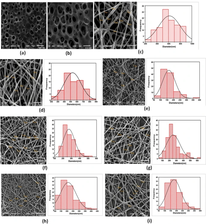
SEM micrographs of (a) PLA cast film, (b) VEN-PLA cast film, (c) 10% PLA NFs, (d) VEN-10% PLA NFs (F1), (e) 10% PCL NFs, (f) 14% PCL NFs, (g) VEN-14% PCL NFs(F2) (h) 10% PLA/PCL composite NFs, and (i) VEN-10% PLA/PCL composite NFs (F3). Image Credit: Hashem, H.M et al., Scientific Reports
Physicochemical characterization of the nanofibers revealed successful loading of the amorphous drug (VEN) into the PLA/PCL nanofibers. The in vitro release of nanofibers showed a 30% burst release of VEN within 0.5 hours, followed by a sustained release of 80% VEN in 96 hours, showing a non-Fickian diffusion mechanism.
Finally, cytotoxicity profiles (IC50) were evaluated in oral epithelial cells for VEN alone, VEN-nanofibers, and nanofibers alone, where the values were 217.55, 250.62, and 440.48 micrograms per milliliters, respectively. Cell toxicity and histopathology studies demonstrated the preservation of the mucosal architecture and preclinical safety of VEN drugs.
Drug Delivery Systems for Controlled Release of VEN
Given the challenges posed by the blood-brain barrier (BBB), treating diseases related to the central nervous system, such as major depression, has become a primary challenge. This barrier prevents the passage of therapeutic molecules with large molecular sizes and low molecular weights, with the exception of small molecules with lipophilic properties.
VEN is a commonly used medication for treating major depression. It is an FDA-approved oral formulation used for pregnant women and adults. VEN is well absorbed from the GIT, with an absorption rate of 92% and an excretion rate of 87% within 48 hours of ingestion. Nanofibers have been utilized as drug delivery systems owing to their facile fabrication and unique properties.
PLA is a renewable material that is widely used in biomedical applications. Its byproducts are biodegradable and extremely safe in the human body and are regarded as suitable hydrophobic media for effective hydrophilic or hydrophobic drug loading.
PCL, which has good drug permeability, a slow degradation rate, and outstanding physicochemical properties for chemical transformations, is also used in medical applications. Consequently, PLA/PCL-blended nanofibers have a synergistic effect owing to their shorter degradation time, superior mechanical properties, and enhanced drug bioactivity.
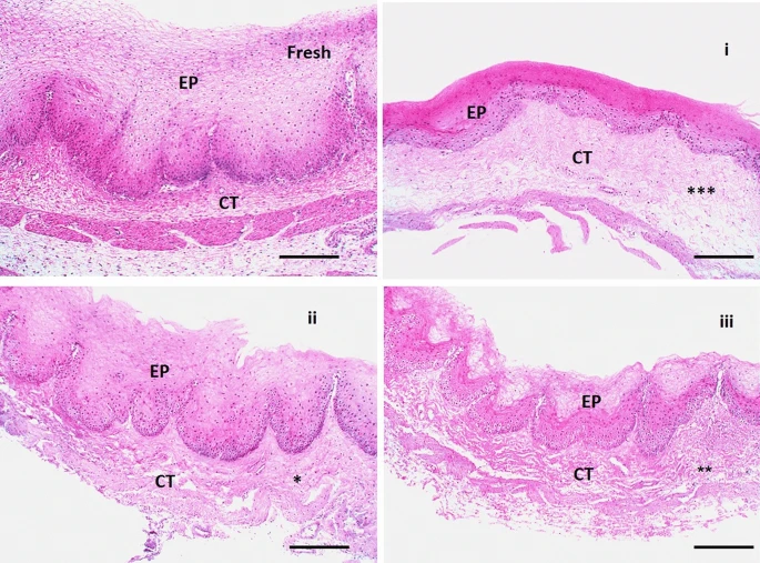
Histopathological pictures of chicken cysts mucosa utilized in ex vivo permeation for a fresh sample as control mucosa, (i) mucosa exposure to VEN solution, (ii) mucosa exposure to plain NFs, and (iii) mucosa exposure to medicated NFs (F3) using H&E stain X:100 bar 100. (*ρ < 0.05, ** ρ < 0.01, ***ρ < 0.001 vs corresponding value of fresh group). Image Credit: Hashem, H.M et al., Scientific Reports
Electrospun Nanofibers for the Sustained Release of VEN
In the present study, biodegradable PLA and PCL polymers were used to prepare nanofibers for buccal delivery of VEN, which helped avoid enzymatic degradation of the drug and hepatic metabolism in the GIT, thereby facilitating controlled drug release.
VEN-loaded nanofibers were fabricated by electrospinning under an applied electrical potential of 20 kilovolts. The solution with biodegradable polymers - PLA, PCL, and PLA/PCL blend along with VEN was electrospun at flow rates of 1.0, 0.6, and 0.3 milliliters per hour, respectively, over a tip-to-collector distance of 15 centimeters.
PLA (10% solution) yielded nanofibers with 400-500 nanometer diameters. The use of a binary solvent system of chloroform: dimethylformamide (80:20) was favorable for producing nanofibers because of its high miscibility, suitable viscosity, low surface tension, and high conductivity.
With an increase in PCL concentration from 10 to 14%, the diameter of the nanofiber gradually increased, suggesting an increase in the electrical force exerted on the jet with increasing PCL concentration, thus enhancing the mass throughput.
However, the diameters of the PCL nanofibers were smaller than those of the PLA nanofibers because the PCL nanofibers had a high crystallinity, degree of molecular orientation, and mechanical properties.
The in vitro release of VEN from nanofibers showed sustained drug release as the drug was dispersed across the carrier and released into the medium. The cumulative amount of drug release indicated rapid drug release from the buffer solution, which was attributed to VEN's high solubility of VEN in water and its ability to make direct contact with the diffusion cellulose membrane.
The PLA/PCL blend demonstrated impactful characteristics and advantages of biopolymer materials that differed from those of individual PLA or PCL biopolymers. Blend formation is facile to spin, allowing for improved material properties of an appropriate blend of individual components (PLA and PCL).
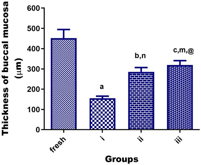
Statistical analysis of the thickness of H&E-stained sections of chicken cyst tissues (as buccal mucosa model) using one-way analysis of variance (ANOVA) followed by the Tukey's for individual comparison of group means to show a significant reduction in fresh mucosa group when exposed to a pure drug of VEN group (i), plain NFs group (ii) or medicated NFs (F3) group (iii). Notes: Different small alphabetical letters mean significant when ρ < 0.05. (aρ < 0.0001; bρ < 0.001; cρ < 0.01 versus corresponding value of a fresh group, n ρ < 0.01 versus corresponding value of VEN group, mρ < 0.05 versus corresponding value of VEN group, @ρ > 0.05 versus corresponding value of plain group). Image Credit: Hashem, H.M et al., Scientific Reports
Conclusion
Overall, PLA/PCL blended nanofibers were presented as a promising and safe controlled-release strategy for the buccal delivery of antidepressant drugs, reducing the frequency of drug administration and the onset of side effects, thereby enhancing drug effectiveness and patient acceptance. In the future, researchers aim to conduct animal studies to make a complete judgment about the developed nanosystem.
Reference
Hashem, H.M., Motawea, A., Kamel, A.H. Bary, E.M.A., Hassan, S.S. (2022) Fabrication and characterization of electrospun nanofibers using biocompatible polymers for the sustained release of venlafaxine. Scientific Reports. https://doi.org/10.1038/s41598-022-22878-7
Disclaimer: The views expressed here are those of the author expressed in their private capacity and do not necessarily represent the views of AZoM.com Limited T/A AZoNetwork the owner and operator of this website. This disclaimer forms part of the Terms and conditions of use of this website.