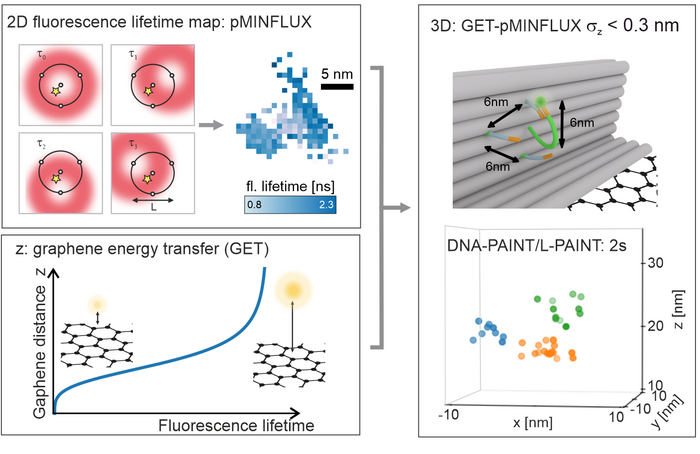
a, pMINFLUX interrogates the position of a fluorophore with multiple spatially displaced doughnut beams and yields 2D fluorescence lifetime images with nanometer precision. b, Graphene provides a measure for the axial distance to graphene. The fluorescence lifetime shortens, the closer a fluorophore is to graphene. c, Combining the lateral information of pMINFLUX with the axial graphene distance information yields 3D localizations. GET-pMINFLUX yields photon efficient localizations with nanometer precision. This enables L-PAINT. The schematic of the DNA origami structure has a DNA-pointer protruding. The fluorophore modified DNA-pointer can transiently to one of three binding sites spaced with 6 nm. Within 2 s this dense structure is with nanometer precision localized in 3D by combining L-PAINT and GET-pMINFLUX. Image Credits: Jonas Zähringer, Fiona Cole, Johann Bohlen Florian Steiner, Izabela Kamińska, Philip Tinnefeld.
This integration, which uses unique characteristics of the 2D material graphene, facilitates an axial accuracy of fewer than 3 angstroms.
The MINFLUX approach questions the position of all molecules by the introduction of a laser focus near the molecule. The fluorescence intensity measured acts as a mean of the distance of the molecule from the laser focus center.
By positioning the center of the laser focus consecutively at varied spots corresponding to the molecule, the exact position of the molecule can be achieved by triangulation. 2D microscopy has already been streamlined using the pMINFLUX (pulsed MINFLUX) developed in the Tinnefeld and Stefani lab in 2020. In this case, the pulsed laser use streamlines lateral super-resolution microscopy and offers access to the fluorescence lifetime.
With the combination of pMINFLUX and graphene, the information of a photon is used in the best possible way. Here, pMINFLUX is synergistically combined with graphene energy transfer (GET) to enable precisions so far unreached for fluorescence microscopy.
Prof. Philip Tinnefeld, Ludwig Maximilian University of Munich
The fluorescence lifetime is based on distance owing to GET. Therefore, a modification in fluorescence lifetime can be transformed from graphene to axial distance. GET-pMINFLUX produces 3D localizations with axial precisions of below 3 Angstroms as a result.
The high precisions achievable with GET-pMINFLUX enable the development of methodologically new approaches to improve super-resolution microscopy which were previously not realizable. Typically, the entire photon budget of a dye is needed to localize a position with 1 nm precision. However, GET-pMINFLUX is so photonefficient that the photon budget can be split among multiple positions and still yield nanometer precise localizations.
Jonas Zähringer, Ludwig Maximilian University of Munich
With DNA nanotechnology, the scientists developed L-PAINT (local PAINT). This is a novel methodology to amplify the speed of super-resolution microscopy. L-PAINT has two binding sequences, unlike DNA-PAINT, that allows super-resolution by unbinding and binding a DNA strand labeled with fluorescent dye.
For this reason, the scientists developed a binding hierarchy in order for the L-PAINT DNA strand to bind longer on one side. This enables the strand’s other end to look for binding sites that come with a high local concentration.
The high local concentration not only increases the speed, but also enables scanning of dense clusters, faster than disturbances due to thermal drift. The combination with GET-pMINFLUX and L-PAINT allows us to study structures and dynamics at the molecular level, which are fundamental for our understanding of cellular biomolecular reactions.
Prof. Philip Tinnefeld, Ludwig Maximilian University of Munich
Journal Reference
Zähringer, J., et al. Combining pMINFLUX, graphene energy transfer and DNA-PAINT for nanometer precise 3D super-resolution microscopy. Light: Science & Applications. https://doi.org/10.1038/s41377-023-01111-8.