 By JanakyReviewed by Lexie CornerUpdated on Aug 1 2024
Atomic Force Microscope (AFM) is an advanced tool widely used for producing highly detailed surface plots at the nanoscale. Originally developed for characterizing nanometer-scaled semiconductor devices, AFMs are now widely used in diverse fields, from material engineering to biophysics, due to their unique analytical capabilities.1

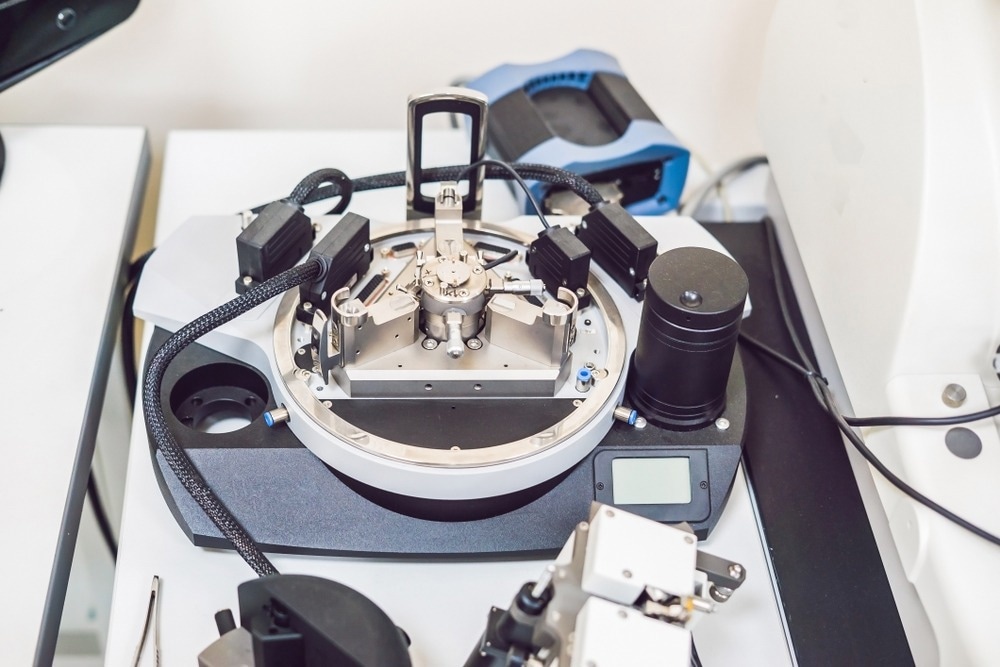
Image Credit: Elizaveta Galitckaia/Shutterstock.com
What are AFMs?
An AFM is an instrument used for mapping the atomic-scale topography of a surface by utilizing the repulsive electronic forces between the surface and the tip of a microscope probe as it moves above the surface.2
AFMs (classified under scanning tunneling microscopes) differ from conventional microscopes, which generate images by focusing light or electrons on the sample surface. Instead, AFMs physically feel the sample surface with a sharp probe, generating accurate three-dimensional topographical representations with nanoscale resolution.1,3 This resolution allows AFMs to image the arrangement of atoms in a sample or observe the structure of individual molecules.
AFMs comprise several components that work together to produce detailed surface plots.4
- Probe: A sharp tip at the free end of a cantilever that is used to scan the surface.
- Cantilever: A flexible arm that holds the probe and bends in response to surface variations.
- Laser: A beam directed at the cantilever, which reflects off its surface.
- Photodiode: A position-sensitive photodetector that detects the laser's movement as the cantilever bends.
How does an AFM work?
An AFM works by raster-scanning a probe line by line across the surface of a sample. As the probe moves, height variations cause the cantilever to bend, altering the deflection of a laser beam aimed at the cantilever, leading to detailed imaging of the sample.2
A laser beam deflection system, commonly used in most AFMs, involves reflecting the laser beam off the back of the cantilever onto the detector.5 A position-sensitive photodiode detects the reflected laser beam, capturing movements to generate a detailed topographical map of the sample surface.
AFM tips and cantilevers are typically micro-fabricated from silicon (Si) or silicon nitride (Si₃N₄), with tip radii typically ranging between a few to tens of nanometers.6 The forces between the tip and the sample impact AFM imaging. The force is not measured directly but is calculated by measuring the deflection of the cantilever, knowing its stiffness.
AFMs use a feedback loop with laser deflection to keep the tip-sample distance constant. As the tip interacts with the surface, the laser spot on the photodetector is employed in the feedback loop to monitor the surface for imaging and measurement.7 This precise control allows AFM to produce highly detailed surface plots, providing unparalleled insights into the intricate surface structures of various materials.
Different AFM Modes
AFM functions in multiple modes to support a range of measurement types.5
- Contact Mode: The probe maintains continuous contact with the surface, allowing high-resolution imaging but potentially damaging softer samples due to constant pressure.5
- Tapping Mode: The probe oscillates close to the sample surface, making brief contact at the lowest point of its oscillation cycle. This mode minimizes potential damage to delicate samples while still delivering high-resolution images.8
- Non-Contact Mode: The probe stays just above the surface, detecting van der Waals forces without making physical contact. This method measures changes in the cantilever's resonant frequency or amplitude due to interaction forces, and it is suitable for analyzing soft or fragile surfaces without inflicting any damage.9
This adaptability makes AFM a powerful tool for exploring the intricate surface structures of a wide range of materials.
Applications of AFM
AFM is most extensively used for its ability to create highly detailed three-dimensional topographical maps with nanoscale resolution. This capability is crucial in semiconductor science and technology, where precise surface imaging is necessary for quality control and imaging of silicon-integrated circuits. AFM has been instrumental in characterizing graphene composite materials, providing insights into their structural integrity and potential applications.10
AFM also facilitates nanomanipulation, allowing researchers to move, bend, or alter nanoparticles with atomic-level accuracy.11 This application is particularly valuable in solid-state physics, where AFM can identify atoms on the surface, evaluate interactions between specific atoms and their neighbors, and study changes in physical properties resulting from atomic rearrangements. The precision supports advanced material engineering and nanotechnology development.
Advanced AFM modes have been developed to provide quantitative data on various material properties, including friction coefficients, electrical forces, capacitance, magnetic forces, conductivity, viscoelasticity, surface potential, and resistance.5 This offers comprehensive insights that correlate surface topology with material properties.
Additionally, this capability makes AFM an essential tool in tribology for studying surface and friction interactions, polymer chemistry and physics for evaluating polymer properties, and energy research for characterizing materials used in energy storage and generation.2
Industries like aerospace and automotive use AFM to develop advanced materials and evaluate their mechanical properties.4 In biological research, AFM is also used to distinguish cancer cells from normal cells by stiffness and investigates protein complexes and cell membrane properties.
Advantages and Disadvantages of AFM
AFM offers several benefits, including the ability to operate in diverse environments such as vacuums, air, and liquids, eliminating the need for a vacuum. This versatility facilitates easy sample preparation and ensures accurate measurements of sample sizes.
AFM can generate detailed 3D images and provide data on physical characteristics, making it ideal for studying both living and nonliving samples, regardless of surface roughness. It is especially valuable in dynamic environments, such as Cellular and molecular biology research, where it captures real-time changes in surface properties and interactions.
Whether assessing material textures, investigating biological specimens, or analyzing surfaces under various conditions, AFM delivers precise and comprehensive insights.
Despite its advantages, AFM has notable limitations. It can scan only a single nanosized area at a time, typically around 150x150 nm, which can be restrictive for larger samples. The relatively low scanning speed can lead to thermal drift, potentially affecting measurement accuracy.4
An article in Surface Topography: Metrology and Properties, investigates how the AFM tip and the sample are prone to damage during detection, especially when scanning narrow valleys, which may compromise results.
Additionally, AFM tips can wear out and require replacement, and the instrument has limitations in magnification and vertical range, which can restrict the observation of certain features in extreme detail.
These drawbacks highlight the need for careful consideration and calibration when using AFM in various applications.
Discover More: How to Achieve Nanomechanical Characterization with AFM
References and Further Reading
- Francis, LW., Lewis, PD., Wright, CJ., Conlan, RS. (2010). Atomic force microscopy comes of age. Biology of the Cell. DOI: 10.1042/BC20090127
- Merriam-Webster. (n.d). Atomic Force Microscope. [Online] Merriam-Webster. Available at: https://www.merriam-webster.com/dictionary/
- Bruker. (2024). High-Resolution Atomic Force Microscopes for Challenging Research. [Online] Bruker. Available at: https://www.bruker.com/en/products-and-solutions/microscopes/materials-afm/
- Mokobi, F. (2023). Atomic Force Microscope: Principle, Parts, Uses. [Online] Microbe Notes. Available at: https://microbenotes.com/atomic-force-microscope-afm/
- Nanoscience Instruments. (2024). What is Atomic Force Microscopy? [Online]. Nanoscience Instruments. Available at: https://www.nanoscience.com/techniques/atomic-force-microscopy/
- Kitazawa, M., Shiotani, K., Toda, A. (2003). Batch fabrication of sharpened silicon nitride tips. Japanese journal of applied physics. DOI: 10.1143/JJAP.42.4844
- Giessibl, F. J. (2003). Advances in atomic force microscopy. Reviews of modern physics. DOI: 10.1103/RevModPhys.75.949
- Oxford Instruments. (2024). AFM: Exploring Tapping Mode and AM-FM. [Online] Oxford Instruments. Available at: https://afm.oxinst.com/outreach/tapping-mode-for-afm-am-fm
- Zurich Instruments. (2024). Non-Contact Atomic Force Microscopy (NC-AFM). [Online] Zurich Instruments. Available at: https://www.zhinst.com/others/en/applications/scanning-probe-microscopy/non-contact-atomic-force-microscopy-nc-afm/
- Humaizi, A. (2024). Atomic Force Microscopy (AFM) for graphene characterization. [Online]. Medium. Available at: https://medium.com/@maizi5469/atomic-force-microscopy-afm-for-graphene-characterization-a9106b6d97b6
- Rubio‐Sierra, FJ., Heckl, WM., Stark, RW. (2005). Nanomanipulation by atomic force microscopy. Advanced Engineering Materials. DOI: 10.1002/adem.200400174
Disclaimer: The views expressed here are those of the author expressed in their private capacity and do not necessarily represent the views of AZoM.com Limited T/A AZoNetwork the owner and operator of this website. This disclaimer forms part of the Terms and conditions of use of this website.