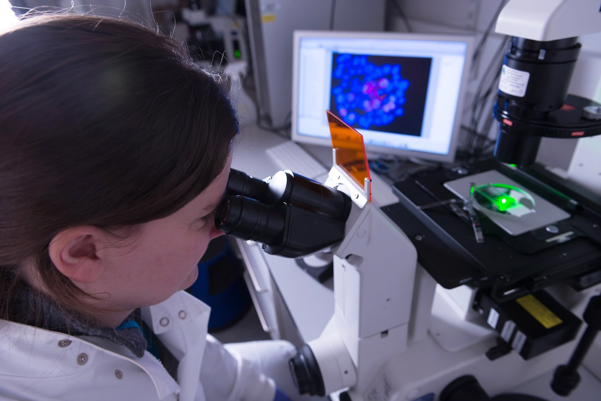
In this interview, Pedro Machado shares expertise on electron microscopy, detailing sample preparation methods and the critical role of multimodal imaging in biological studies.
Can you briefly explain what multimodal biological electron microscopy is and why it is important for biological research?
Multimodal biological electron microscopy involves studying the same material using multiple microscopy techniques. This approach is crucial as it allows us to gather diverse types of data from the same area of interest.
For instance, we can collect structural, molecular, and compositional information at various scales, providing a comprehensive view of the sample. This holistic perspective is particularly valuable in biological research, where understanding the complexity of a sample often necessitates examining it from multiple angles.
What are some of the main challenges you encounter when using electron microscopy for biological samples?
One of the most significant issues is the sensitivity of biological samples. Since these samples are readily destroyed during the imaging process, they must be prepped and handled with care.
We also encounter sample size and mounting issues, particularly when dealing with multimodal imaging. Ensuring that the sample stays intact and appropriately orientated during the various imaging steps is critical for getting accurate findings.
There are two key sample preparation techniques: room temperature fixation and cryo immobilization. What are the advantages and disadvantages of each?
Room temperature fixing often uses chemical agents to preserve the sample's structure, allowing it to be observed in a relatively stable form. The advantage of this method is its familiarity and effectiveness with a wide variety of samples. However, these chemical treatments can introduce artifacts or distortions.
Cryo immobilization, on the other hand, involves rapidly freezing the sample to preserve its original state as accurately as possible. This technique is effective in preventing ice crystal formation, which could damage the sample. The downside is that it requires more specialized equipment and procedures, and the samples must be kept at extremely low temperatures throughout the process.
How do fixation and hardening processes differ between room temperature and cryo preparations, and why are these steps crucial for electron microscopy?
In room temperature preparations, fixation is commonly accomplished using chemicals such as formaldehyde or glutaraldehyde, which cross-link proteins and other biological components to stabilize the sample. The sample is then hardened with resins to make it simpler to section for microscopy.
Fixation in cryo preparations involves fast freezing the sample, generally in liquid nitrogen, which preserves the sample without the need for chemicals. The cryo-hardening method includes freeze substitution, which replaces the water in the sample with an organic solvent at low temperatures. These procedures are critical because they influence the quality of the finished preservation. Proper fixation and hardening guarantee that the material retains its structure and allows us to slice it into very thin slices for imaging.

Image Credit: Connect Images - Curated/Shutterstock.com
What methods are used for shaping and sectioning samples for electron microscopy, and how do these methods impact the imaging quality?
Microtomes are tools intended to cut exceedingly tiny slices of a sample and are often used for shaping and sectioning. For room temperature samples, this occurs after the sample has been immersed in resin, which provides essential support. In cryopreparations, we employ cryomicrotomes, which operate at very low temperatures to retain the material frozen during sectioning.
The quality of these sections directly impacts the resulting images. If the sections are too thick or uneven, the images may suffer from poor resolution and contrast. In contrast, thin, well-prepared sections produce clear, high-resolution images, which are essential for accurate analysis.
How do energy dispersive spectroscopy (EDS) and backscattered electron (BEX) imaging techniques contribute to the understanding of sample composition and structure?
EDS and BEX are effective methods for determining the elemental composition of materials. EDS enables us to identify and measure the various elements in a sample by detecting the X-rays released when it is hit with electrons. This approach is particularly effective for determining how various stains and contrast ants are spread throughout the sample.
BEX imaging offers compositional contrast by detecting electrons scattered back from the material combined with the elemental information. This approach focuses on fast imaging, enabling us to immediately see the locations of various components or compounds inside the sample. Together, these procedures provide a complete picture of the sample's composition and assist us in optimizing our preparation methods.
Can you share some specific examples or case studies where EDS and BEX were particularly useful in your research?
One notable example is when we utilized EDS to compare various labeling methods for parasitic cells such as Leishmania. By analyzing the elemental makeup of cells prepared with various stains, we were able to determine why certain methods offered higher contrast than others.
For example, we discovered surprising findings with uranyl acetate stains, where the uranium signal did not correspond with the applied concentration, prompting us to reconsider and improve our staining processes.
Another example is BEX imaging of a birch seed, which allowed us to swiftly map the distribution of components across the sample. This enabled us to pinpoint areas of interest for additional investigation, making the whole process more efficient and focused.
How has the integration of different imaging modalities, such as Raman imaging with SEM, advanced the field of biological electron microscopy?
Integrating diverse imaging modalities, such as Raman imaging and SEM, has revolutionized biological electron microscopy. Raman imaging offers molecular bonding information and chemical identification, which, when paired with SEM structural and compositional data, provides a far more complete picture of the material.
For example, in our study of hydroxyapatite implants, we employed a multimodal method to examine the interface between the implant material and bone tissue. By integrating Raman and SEM data, we were able to detect new bone development locations and understand their chemical makeup, which was critical for our investigation.
In your experience, what are some best practices for optimizing sample preparation to achieve high-quality multimodal imaging results?
One of the best practices is to meticulously plan the sample preparation with the final objectives in mind. This involves considering the specific requirements of each imaging modality from the very beginning. For instance, if both electron microscopy and Raman imaging are to be used, it is crucial to ensure that the preparation process does not compromise either technique, such as avoiding coatings that could distort Raman signals.
Another essential practice is to maintain consistent sample orientation and mounting throughout the imaging process. This consistency is crucial for correlative studies, where images from multiple modalities must be accurately compared. Additionally, minimizing electron radiation during imaging is vital, especially for delicate biological samples, to prevent damage and ensure high-quality results.
What future developments or innovations do you foresee in the field of multimodal biological electron microscopy that could further enhance sample preparation and imaging techniques?
Looking ahead, I expect to see advancements in detector technology that will enable increasingly sensitive and high-resolution imaging across various modalities. Additionally, AI and machine learning could be used to help enhance image analysis by predicting the optimal sample preparation procedures based on the sample type and intended imaging outcomes.
Another promising development might be the improvement of cryo-preparation procedures, making them more accessible and simpler to use. This would enable improved preservation of biological samples in their native conditions, resulting in more accurate and detailed imaging.
In addition, advances in software for correlating data from several modalities may simplify integrating and comprehending large amounts of information, therefore improving our knowledge of biological systems.

This information has been sourced, reviewed and adapted from materials provided by Oxford Instruments.
For more information on this source, please visit Oxford Instruments.
Disclaimer: The views expressed here are those of the interviewee and do not necessarily represent the views of AZoM.com Limited (T/A) AZoNetwork, the owner and operator of this website. This disclaimer forms part of the Terms and Conditions of use of this website.