Atomic Force Microscopy is a highly valuable characterization tool. Typically used for micro/nanostructured coatings, this flexible technique can obtain high-resolution nanoscale images and study local sites in air or liquid surroundings.1 Specialist Steven Marsden talks us through everything AFM, from the basics to the exciting developments on the horizon.
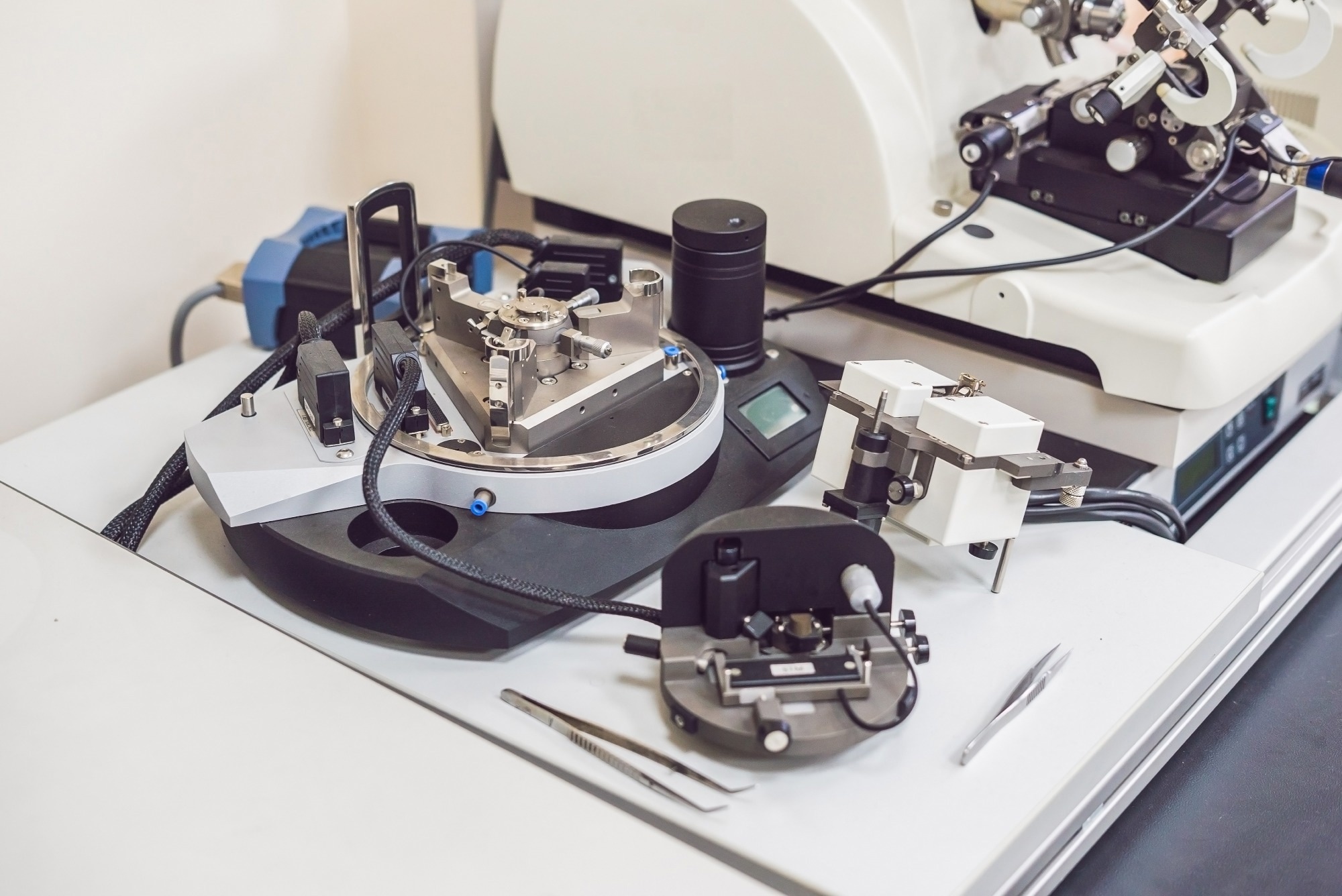 Image Credit: Elizaveta Galitckaia/Shutterstock.com
Atomic Force Microscopy Explained
First invented in 1985 by IBM in Zurich, Atomic Force Microscopy (AFM) is a scanning probe technique for imaging. It involves a nanoscopic tip attached to a microscopic, flexible cantilever, which is raster scanned over a surface.2
You're essentially feeling out the contours of a surface as you scan. The tip is so sharp it can fit in and around even single molecules, so you can get very high lateral resolution as well.
Steven Marsden, Senior AFM Specialist at the University of Manchester, Stopford Building
As the tip scans, a laser bounces off the back of the cantilever onto a position detector, which can be translated into surface height measurements. A very small reflection of the cantilever causes a much larger deflection, so tiny changes in height can be monitored, down to about half a nanometer resolution. Lateral resolution routinely goes down to about five nanometers.
The resolution of AFM images hinges entirely on the size of the needle, and with an ultrasharp tip, lateral resolution of just one nanometer can be achieved.
The Bruker tips below are a standard, more basic size for initial scanning. Holding them to the light and squinting, you can just about glimpse a shimmer of the microscopic needle.
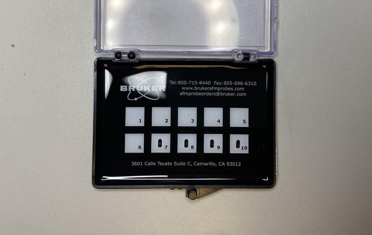 Image Credit: Frances Briggs/AZoNetwork
These days, algorithms and computing techniques can elevate resolution even further, down to 0.1 nm.
This works well in certain circumstances, say if you can take multiple images quite quickly of the same sample, you can get the surface detail smaller than the tip. The limiting factor is how sharp or blunt the tip is. If you have a blunt tip, and your sample is sharp, you’ll just end up with images of the tip.
Where you have a gap in a sample, you’re more likely to have noise there than if you are constantly in contact with the surface. If you have lots of images, you can use probability to describe the gap you can’t see physically, by sensing it, in a way, indirectly. These probability maps generated by algorithms are what can improve the resolution, comparable to electron microscopy.
Steven Marsden, Senior AFM Specialist at the University of Manchester, Stopford Building
Depending on the sample, AFM can be done in air or in liquid. Steve Marsden describes the in-air atomic force microscope in his imaging suite as the workhorse system.
Whether imaging in air or in a liquid there are different advantages and disadvantages. In air, you have issues with water layers, so samples have to be really dry. It’s sometimes easier though because you don’t have things bashing into the tip and so on.
Steven Marsden, Senior AFM Specialist at the University of Manchester, Stopford Building
There are various ways to reduce noise in a sample. This can include conducting imaging in closed environments, as shown below in Steven's protected, insulated mini AFM room, or simply on a specialized desk that absorbs shocks in the environment, such as building noise.
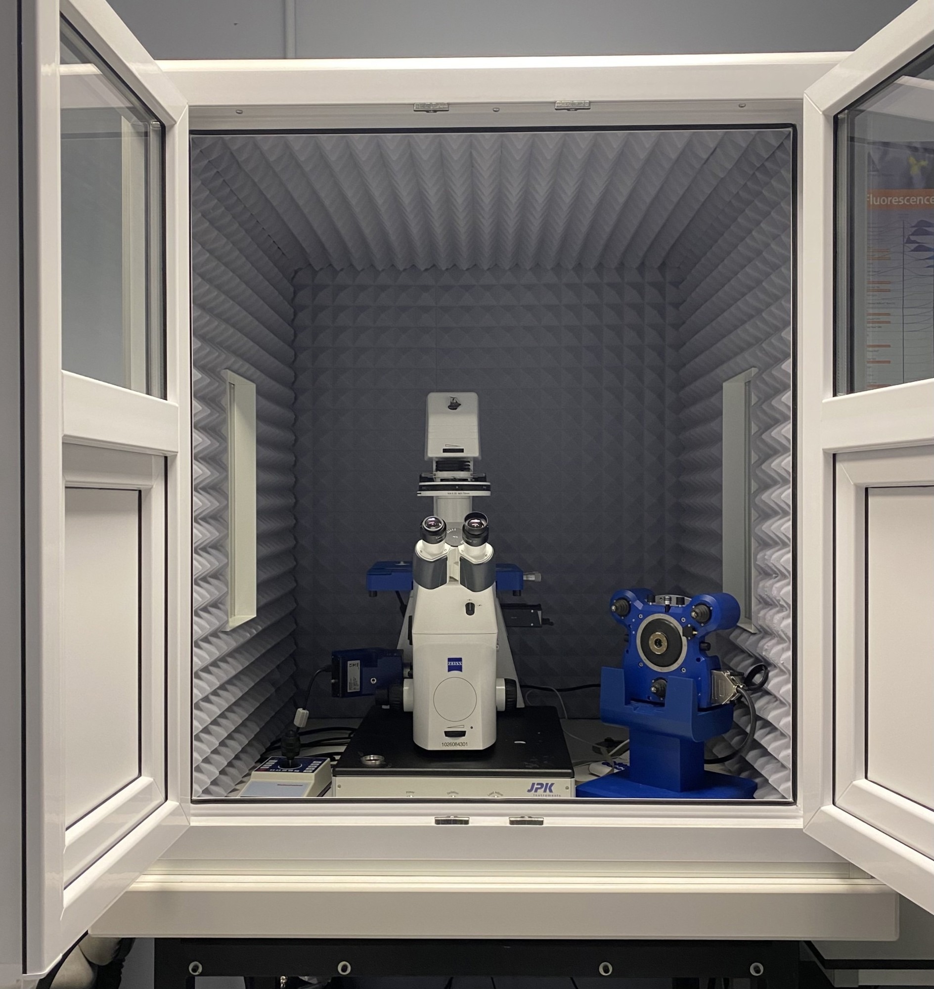 Image Credit: Frances Briggs/AZoNetwork
Challenges and How to Solve Them
Usually, the biggest challenges include sample stability. If the sample is really wobbly or if it is loosely attached you can have problems imaging that. Contamination is also a problem. In solution you can get bits floating around on the surface, so if you have any buffers with salt all you will see is just mountains of salt!
With contamination it’s hard to distinguish between your sample and contamination. That’s why you need lots of controls, with and without your sample, building up different components one at a time to understand the full picture of what your sample should look like.
Steven Marsden, Senior AFM Specialist at the University of Manchester, Stopford Building
However, technology has moved on since its invention in 1985, and different AFM modes can be used to evade sample stability issues and reduce damage to materials as they are imaged. The original mode of AFM imaging, with the tip always in contact, can destroy the surface. Using a tapping mode, for example, means the tip is not always in contact with the surface, making it less destructive.
Another mode uses forces to measure a surface. As well as being able to image with AFM, measuring interactions with the surface can provide information on the mechanical and potentially electrostatic forces between the tip and the surface, painting an even more detailed picture of a sample's topology.
Combining the known spring constant of the cantilever with the data from force microscopy can reveal the stiffness of a sample or even the force required to pull away from the surface. According to Steven, the latest imaging techniques combine the ability to measure forces with imaging.
You get all that data in each image pixel. And it also means, because you’re measuring the force of interaction you have more control over it, so you can have the softest force as you scan, making the technique kinder on the sample. You can use algorithms in the software to adjust all parameters from all this information you have.
Bruker uses Scan Asyst to help resolve those parameters to give you the best surface contact and imaging quality as you scan. These parameters change on the fly, so now you don’t have to worry about recording parameters as you scan. It makes AFM a lot easier to do.
Steven Marsden, Senior AFM Specialist at the University of Manchester, Stopford Building
Download your PDF now!
What is to Come in AFM?
Since its invention, AFM has become a valuable tool for characterization, as a technique that is quick and easy. It provides researchers with a way to see their samples in their native state, without having to stain them, or tag them, or even conduct imaging in a vacuum.
With a biological background, Steven is interested in how AFM might be used as a diagnostic tool, as well as in its use in nanomechanics.
In cell biology, nanomechanics involves the investigation of the environment around cells and how changes to external forces can have an impact on a molecular level. Different forces exerted on a cell during development can mean the difference between a new brain cell and a bone cell. AFM works well in monitoring these changes as it can provide data on the forces in the environment and simultaneously image the changes that are occurring.3
Using AFM in combination with other techniques is another interesting evolution. Using it in tandem with raman spectra can give you higher spatial resolution and chemical characterization of a surface.
That could be interesting in biology because you could image a tissue, lets say, and then get all this spectra, ask what has happened to the patient who gave us this tissue. Perhaps AI could mine this and use a database to find out the tissue’s chemical composition is indicative of tumor growth, so it could even be a diagnostic tool if it were used like this.
Steven Marsden, Senior AFM Specialist at the University of Manchester, Stopford Building
 Image Credit: Frances Briggs/AZoNetwork
Four decades after its invention, Atomic Force Microscopy remains one of the most adaptable tools in modern science. Once a niche imaging method, it continues to edge toward diagnostic and analytical frontiers, probing the mechanics of cells, mapping materials in real time, and coupling with spectroscopic and AI techniques to interpret chemistry at the nanoscale. As resolution improves and automation expands, AFM is being remodeled as intelligent sensing.
About Steven Marsden
Steven Marsden is the Senior Biological Imaging Specialist at the University of Manchester. He works with a range of equipment, from AFM to SEM, and has a depth of knowledge in all things nano-imaging.
References and Further Reading
- Aliofkhazraei, M. and Ali N., (2014) FM Applications in Micro/Nanostructured Coatings, Comprehensive Materials Processing, (7) 191-241.https://doi.org/10.1016/B978-0-08-096532-1.00712-3
- Vahabi S, Nazemi Salman B, Javanmard A. (2013) Atomic force microscopy application in biological research: a review study. Iranian Journal of Medical Sciences. https://pmc.ncbi.nlm.nih.gov/articles/PMC3700051/
- Kilpatrick, J. I., Revenko, I., Rodriguez, B.J. (2015) Nanomechanics of Cells and Biomaterials Studied by Atomic Force Microscopy, Advanced Healthcare Materials. https://doi.org/10.1002/adhm.201500229,