Feb 14 2019
Magnetic nanoparticles are being extensively used in tissue bioengineering and cell imaging, yet, what happens to them inside stem cells, in the long run, is still not documented. Scientists from CNRS, the Sorbonne Université, and universities Paris Diderot and Paris 13, have demonstrated considerable degradation of these nanoparticles, followed in some cases by the cells “re-magnetizing.” This occurrence is the sign of biosynthesis of new magnetic nanoparticles from iron discharged in the intracellular medium by the degradation of the initial nanoparticles. Reported in PNAS on February 11, 2019, this research may elucidate the presence of “natural” magnetism in human cells, and help scientists to visualize new tools for nanomedicine, because of this magnetism formed by the cells themselves.
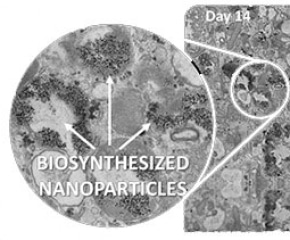 © Laboratory MSC (CNRS/University of Paris Diderot)
Magnetic nanoparticles play a crucial role in present-day's nanomedicine: they serve as imaging diagnosis agents, drug targeting agents, thermal anti-cancer agents, and tissue engineering agents. The question of their destiny in cells, after they have fulfilled their therapeutic role, was not properly understood.
To track the passage of these nanoparticles in cells, scientists at the Laboratoire Matière et Systèmes Complexes (CNRS/Université Paris Diderot) and the Laboratoire de Recherche Vasculaire Translationnelle (INSERM/Université Paris Diderot/Université Paris 13), in partnership with researchers from Sorbonne Université have formulated a novel approach to nanomagnetism in living systems: first they integrated magnetic nanoparticles in vitro in human stem cells. They then left them to separate and form for one month, to track them long term in the intracellular environment and to check their alterations.
By tracking the “magnetic fingerprint” of these nanoparticles in the cells, the scientists have demonstrated that they were first being degraded (cell magnetization declines) and discharging iron into the intracellular environment. Subsequently, this “free” iron was stored in non-magnetic form in ferritin, the protein accountable for storing iron, or served as a foundation for the biosynthesis of new magnetic nanoparticles within the cell.
This occurrence is said to take place in some bacteria, but a biosynthesis like this had never been witnessed in mammalian cells. This could elucidate the existence of magnetic crystals in humans, seen in the cells of diverse organs, mainly the brain. Furthermore, this iron storage in magnetic form could also be a path for the cell to “detoxify” over the long term to balance out extra iron. From the standpoint of nanomedicine, this biosynthesis paves the way to the prospect of purely biological magnetic marking in cells.