Confocal microscopy is an optical imaging technique that employs a point light source (laser), which is directed onto the sample. The same objective usually collects the reflected (fluorescence) light, which is focused into a pinhole in front of the detector. This is made to make sure that only light from the image focal plane reaches the detector, which considerably increases image contrast; resolution can also be increased with appropriate selection of pinhole size.
Confocal Raman Imaging
Particularly in confocal Raman imaging, the acquisition time for one Raman spectrum is very important because it influences the acquisition time of the image which normally includes thousands of Raman spectra. This article illustrates how the use of a spectroscopic EMCCD as the detector can considerably reduce the acquisition time down to a few milliseconds per spectrum, while enhancing sensitivity.
In confocal Raman imaging, as in WITec’s alpha300R system, unique filters are utilized to restrain the reflected laser light while allowing the Raman scattered light to be detected with a combination of CCD camera and spectrometer. In order to acquire an image, thousands of spectra are obtained in an extremely short time with less than 100ms integration time per spectrum. However, since the Raman scattering cross section is small and the excitation power is restricted to a few milliwatts, it becomes challenging to enhance the overall sensitivity of a confocal Raman system.
Improving Overall Sensitivity of Confocal Raman Imaging
To improve the overall sensitivity of confocal Raman imaging, the first step is to enhance the collected Raman signal. This can be achieved by optimizing the throughput of the spectrometer and microscope and also by utilizing an objective with a high numerical aperture. The confocal arrangement helps in reducing unwanted background signal and further improves signal-to-noise ratio. To this end, a number of factors have to be considered to improve the overall sensitivity. These factors include dark noise, readout noise, and selection of an appropriate detector.
Dark Noise
Dark noise usually occurs on account of thermally generated carriers in the CCD, which can be significantly reduced by cooling the CCD. Since a good CCD exhibits a thermal dark current of less than 0.01 electrons/pixel/second at -60°C, cooling below -60°C is not needed for integration times of a few seconds.
Readout Noise
Camera manufacturers specify the readout noise, which is given in electrons. Standard values are 5-10 electrons for a 50kHz readout rate to approximately 30 electrons at 2.5MHz readout rate. When the readout noise surpasses the photon shot noise, the signal is believed to be readout noise limited. In typical spectroscopic experiments, the integration time is increased to obtain sufficient signal so as to ensure that the signal is shot noise limited again. However, in confocal Raman microscopy, this is not always possible.
Detector
Choosing an appropriate detector with the highest sensitivity is very important. Dark noise and the readout noise are the main sources of noise of the detector. The aim must be to remove all noise sources so that only the noise of the photon shot remains.
For integration times of below 100ms, such as in the confocal Raman set-up, the dark current is entirely negligible. Readout noise, which is created while changing the collected electrons into digital counts, is restricted by the speed of the readout process and the quality of the CCD´s readout amplifier.
EMCCD
An electron multiplying CCD (EMCCD) is a standard back-illuminated CCD with an extra readout register which is driven with a higher clock voltage than a normal CCD readout register. Owing to this high clock voltage, an electron multiplication via impact ionization is obtained by amplifying the signal up to 1000 times. With this arrangement, it is possible to increase the signal higher than the readout noise so that the signal-to-noise ratio is always restricted by the Poisson noise of the signal, even if an extremely fast readout amplifier is employed. For instance, a 1600 x 200 pixel EMCCD with a 2.5MHz readout amplifier can be read out in just 2.3ms.
For higher signals, wherein the intensity of signal is not readout limited, the excess noise factor of the EM process reduces the EMCCD’s signal-to-noise ratio to less than that of a standard CCD. In such cases, the EM register can be switched off and the “normal” readout register is utilized.
Raman Imaging with EMCCD
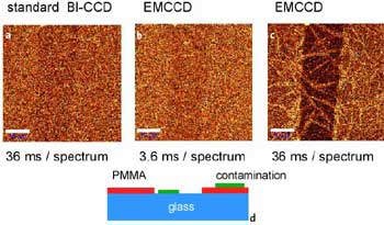
Figure 1. a-c: Confocal Raman images of a 7.1 nm thin PMMA layer on glass obtained in the CH2 stretching band around 3000 / cm. Scale bar: 10 µm. Fig. 1d: Schematic of the sample 30 x 50 µm, 100 x 80 pixel = 8000 spectra, 110 ms/spectrum.
The middle image i.e. Figure 1b depicts the same part of the sample imaged with an EMCCD with a gain of approximately 250. The image demonstrates almost the same signal-to-noise ratio, but now the integration time was only 3.6ms, which is 10X faster than Figure 1a. For Figure 1a, the complete image acquisition took 25 minutes while it took only 3.4 minutes for Figure 1b. The EMCCD was used to take Figure 1c, but now with the same integration time as in Figure 1a. One can obviously see the scratch and contamination by way of a needle-like structure across the glass surface and PMMA. Figure 1d illustrates a sketch of the sample.
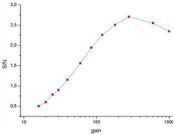
Figure 2. Comparison of confocal Raman images acquired with different settings of the EMCCD gain.
In Figure 2, the signal-to-noise ratio of the signal from the CH2 stretching band of the PMMA is plotted against the EMCCD gain.
Conclusion
Thus, with the help of an EMCCD camera, speed and detection efficiency can be greatly increased, particularly for the short integration times required with a confocal Raman microscope. The distribution of a 7.1 nm PMMA can be easily identified with an integration time of 7ms per spectrum, which considerably reduced the overall acquisition time to 5.4 minutes for a 200 x 200 spectra confocal Raman image.
For larger signals, the electron multiplying circuit can be switched off and all properties of a normal back-illuminated CCD are maintained., while for extremely small signals the EMCCD can greatly enhance the signal-to-noise ratio by a factor of 5 -10.

This information has been sourced, reviewed and adapted from materials provided by WITec GmbH.
For more information on this source, please visit WITec GmbH.