Feb 18 2011
Research involving selective irradiation of a human skin tissue model is improving how scientists determine the overall effects of low doses of ionizing radiation such as might be received during certain medical procedures or occupational exposures.
Scientists at Washington State University-Tri Cities and the Pacific Northwest National Laboratory are modeling electron energy deposition patterns as produced by the electron microbeam developed at PNNL to determine how it may be used in the study of more complex biological systems.
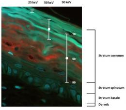
 Microscope image of a vertical slice through a skin-tissue model showing the range of penetration depths calculated for 25-, 50-, and 90-keV electron beams. The centers of the bars mark the depths at which half of the electrons are expected to stop. Approximate widths of the three layers in the epidermis are indicated.
Cells growing as a monolayer in a Petri dish are frequently used for determining responses to environmental insults such as radiation. However, this model does not include cell-cell and cell-matrix interactions critical to maintaining tissue homeostasis. Using a more realistic and complete tissue model is critical to the development of a mechanistic understanding of the cellular radiation responses that occur in vivo.
In this study, the WSU-TC and PNNL scientists used an artificial skin model made of normal human epidermal skin cells and connective tissue cells called fibroblasts. The advantage of the model is that it has a well-defined cellular composition that scientists can modify to gain a fundamental understanding of how different cell types interact following irradiation. This understanding will help development of biologically based risk models and help ensure that radiation protection standards are adequate and appropriate.
In a series of recent papers, fluorescent and confocal microscopy images have been used to characterize the detailed cellular morphology of the skin tissue model. Using these images as a guide, Monte Carlo simulations have been performed to establish the energy deposition patterns on the microscopic scale. The computer simulations determined the feasibility of selectively irradiating only the epidermal layer using the PNNL-developed electron microbeam. Microbeams provide a convenient way to investigate radiation-induced bystander effects. Bystander effects are responses in unirradiated cells that are triggered by signals received from irradiated neighboring cells.
Results of the computer simulation suggest that the skin-tissue model epidermis can be irradiated without significant exposure to the dermal layer. This is because of the energy dependence of the electron microbeam's penetration of the skin sample. The result is a more realistic radiation energy deposition scenario.
Now that the scientists are confident that selective irradiation of the epidermis of skin tissue is feasible using the PNNL electron microbeam, they will begin using this device to understand the role of particular cell types in the radiation-induced response.
Source: http://www.pnl.gov/