From missing particles to grain boundaries, the review shows how paired imaging and AI tools could make hidden flaws in self-assembled nanomaterials easier to detect and quantify.
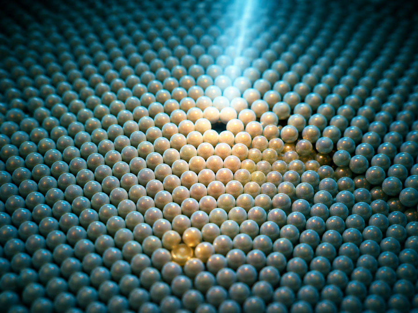
Paper: Nanosphere Self-Assembly Imaging Systems and Defect Detection Algorithms for Self-Assembled Structures: A Review. Image credit: AI-generated image created using ChatGPT/OpenAI
In a recent review article published in the journal Nanomaterials, researchers systematically reviewed imaging systems and defect detection algorithms for self-assembled nanosphere structures, emphasizing integrated approaches that combine advanced microscopy and machine learning to improve quantitative defect inspection and process optimization at the nanoscale.
Nanosphere Assembly & Defects
Self-assembled nanosphere structures serve as powerful bottom-up platforms to fabricate ordered micro- and nanostructures with diverse applications in photonic crystals, sensing, coatings, drug delivery, and nanosphere lithography.
Their functional performance depends critically on structural parameters, such as particle packing density, ordering, interparticle spacing, and the presence of defects, including vacancies, interstitial particles, dislocations, grain boundaries, stacking faults, voids, and cracks.
These imperfections can arise from factors like particle size dispersity and nonequilibrium assembly kinetics, disrupt periodicity, and may compromise device performance. Reliable, quantitative defect inspection is therefore essential for understanding assembly mechanisms, evaluating structural quality, and optimizing fabrication processes.
Although various imaging methods are available, no single modality fully satisfies the requirements for defect characterization due to trade-offs in spatial resolution, field of view, temporal resolution, and operational conditions.
Moreover, traditional image analysis techniques struggle with complex defect morphologies, motivating the integration of advanced imaging with machine learning and deep learning algorithms to support automated, high-throughput, and accurate defect detection in nanosphere assemblies.
Imaging Modalities & Algorithms
The review systematically surveys nanosphere self-assembly structures categorized by dimensionality, zero-, one-, two-, and three-dimensional arrangements, using classical epitaxial growth modes as a conceptual kinetic framework for interpreting resultant architectures and defect formation.
It then comprehensively evaluates major imaging systems, including optical microscopy, electron microscopy, scanning electron microscopy (SEM), transmission electron microscopy (TEM), liquid-phase TEM, atomic force microscopy (AFM), and scanning near-field optical microscopy (SNOM), emphasizing their resolution limits, field of view, in situ capabilities, and compatibility with multimodal correlative approaches. The paper details the advantages and challenges of each modality for nanoscale defect visualization.
A particular focus is placed on emerging integrated imaging workflows that combine large-area rapid screening, high-resolution defect localization, three-dimensional topography, and dynamic process observation under ambient or liquid environments.
On the algorithmic front, the review categorizes defect detection strategies into classical image processing (including thresholding, edge detection, and frequency analysis), machine learning (including feature extraction with statistical classifiers), and deep learning (including end-to-end hierarchical feature learning for segmentation and object detection).
It discusses their respective applicability, robustness, and data requirements, as well as the potential use of weakly supervised and self-supervised learning to reduce annotation burdens in nanosphere assemblies. The authors emphasize the importance of standardized datasets, transferable algorithms, and closed-loop feedback to integrate imaging and analysis into intelligent defect inspection systems.
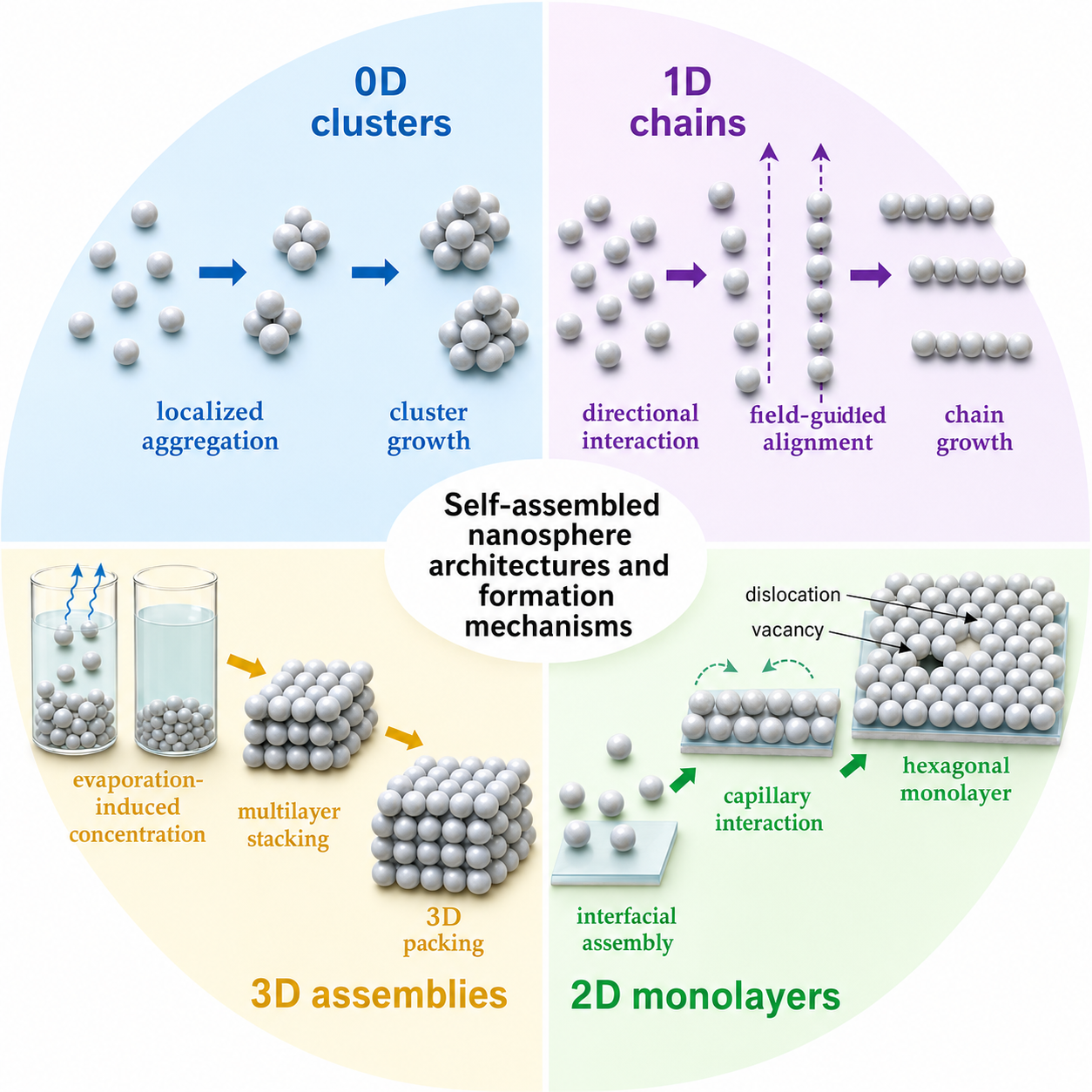
General schematic illustration of representative self-assembled nanosphere architectures and their formation mechanisms. Zero-dimensional clusters formed through a nucleation-dominated pathway, including dispersed nanospheres, localized nucleation, aggregation, and island growth; one-dimensional chain-like assemblies generated by directional interparticle interactions or field-guided alignment; two-dimensional monolayers formed by interfacial assembly and capillary-driven organization into hexagonal close-packed arrays, with typical defects such as vacancies, dislocations, and grain boundaries; three-dimensional assemblies produced by evaporation-driven concentration or sedimentation followed by multilayer stacking and three-dimensional packing.
Defect Detection & Analysis
The review notes that optical microscopy offers rapid, nondestructive, large-area imaging but limited spatial resolution, which is insufficient for nanoscale defect characterization. SEM provides high-resolution surface morphology, while TEM can examine internal structures and, in suitable samples, atomic-scale features, but both may have low throughput, vacuum requirements, and potential beam damage, limiting their use for delicate or dynamic samples.
AFM stands out for its ability to generate three-dimensional topographical maps and to perform dynamic, in situ observation at solid/liquid interfaces, while high-speed AFM has achieved millisecond temporal resolution in liquid-phase studies and may help track nanoparticle assembly and defect evolution.
SNOM complements these with nanoscale optical-field mapping, which is beneficial for studying plasmonic hotspot distributions in metal nanosphere arrays. Correlative microscopy integrating these modalities can help address individual limitations, supporting multiscale structural and functional characterization useful for defect detection and understanding assembly dynamics.
Algorithmic approaches reveal a progression from classical methods, suitable for simple, high-contrast defect detection but sensitive to noise and ill-suited for complex morphologies, to machine learning techniques that incorporate handcrafted features for improved classification, albeit constrained by feature expressiveness.
Deep learning models trained on annotated datasets can improve the segmentation and localization of diverse defect types in complex images. However, they demand large, high-quality, and material-specific datasets, limiting generalizability across different nanosphere compositions or imaging modalities.
The development of self-supervised and weakly supervised learning frameworks shows promise for reducing these data annotation burdens. Furthermore, physics-informed neural networks and domain adaptation strategies could incorporate knowledge of crystal symmetry and assembly kinetics, thereby enhancing interpretability and reliability.
Future Directions & Integration
Self-assembled nanosphere structures offer promising routes to fabricate ordered nanoscale architectures with broad applications, but are inherently prone to defects that may degrade performance.
Effective defect inspection requires combining appropriate imaging modalities, each with trade-offs in resolution, speed, and environment compatibility, with advanced algorithmic analysis ranging from classical image processing to state-of-the-art deep learning methods.
Current challenges include insufficient cross-modality data standardization, limited transferability of learning models across materials and imaging systems, and the need for extensive annotated datasets. Future progress may benefit from multimodal correlative imaging integrated with data-efficient, physics-informed machine learning frameworks.
Such integration, coupled with real-time feedback control, could facilitate automated, quantitative, and adaptive defect inspection workflows. This paradigm may deepen fundamental understanding of assembly mechanisms and help advance scalable, reliable nanomanufacturing of functional self-assembled nanomaterials.
Source:
- Liu Q., Chen Y., et al. (2026). Nanosphere Self-Assembly Imaging Systems and Defect Detection Algorithms for Self-Assembled Structures: A Review. Nanomaterials 16(14):890. DOI: 10.3390/nano16140890, https://www.mdpi.com/2079-4991/16/14/890