Graphene research has been greatly aided by the noninvasive features of optical microscopy over the last decade. This article will discuss the importance of optical microscopy in graphene research, its compatibility with optical graphene visualization, and new developments in this field.
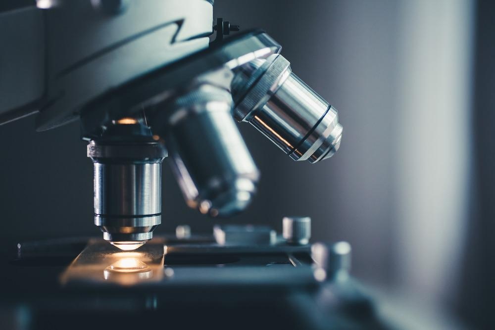
Image Credit: Konstantin Kolosov/Shutterstock.com
Optical Microscopy and the Challenges
In the field of materials characterization, optical microscopy is one of the most commonly used imaging techniques. It is estimated that the diffraction limit of optical light is half of the wavelength, an important consideration in determining the smallest distances that a microscope can resolve.
In the case of visible light, this generally means that objects smaller than 200 nm are difficult to distinguish. As a single layer of graphene can be as thin as 0.34 nm, using optical microscopy to image graphene requires a different approach than simply imaging the object with visible light.
Furthermore, direct optical visualization of graphene on transparent substrates remains difficult due to its low optical contrast of 10%. The local number of graphene layers is difficult to quantify even with advanced setups. Visualizing nanoscale defects in graphene, like cracks, voids, wrinkles, and multilayers, that form during growth or subsequent transfer process, adds another level of difficulty to most device substrates.
New Developments
One method for imaging graphene that does not require the use of electron microscopy is interference reflection microscopy (IRM), which is a label-free optical microscopy technique in which a collimated beam of filtered lamp light propagates through the substrate and is reflected off, with the resultant reflection interference offering optimum contrast for graphene layers.
For example, optical contrast of up to 42% has been reported for monolayer graphene on transparent substrates by researchers. In addition to its high contrast, IRM can clearly reveal nanoscale structures, defects, folding, nanoscale cracks, and wrinkles in graphene, which conventional microscopy cannot. Furthermore, IRM offers high-quality visualization of nanoscale contaminants that correlate well with scanning electron microscopy (SEM) and atomic force microscopy (AFM) results.
Aside from that, wide-field images were recorded in 10 ms snapshots, which is 1000 times faster than SEM and >10,000 times faster than AFM. Furthermore, IRM does not require a vacuum and avoids sample damage from a scanning tip or electron beam.
Surface Plasmon Resonance (SPR), a surface-sensitive optical analysis technique capable of measuring the properties of ultrathin films, is another high-throughput option for imaging graphene. It has been demonstrated that the SPR method can distinguish the thickness of various graphene layers and provide an approximate value of 0.37 nm for the thickness of the CVD-grown monolayer graphene layer, which agrees exactly with the literature value. This method has the advantage of being a quick and low-cost imaging technique.
Optical Imaging of Graphene by Machine Learning
Since the introduction of monolayer graphene, the interpretation of the number of graphene flakes has been based on the color contrast of graphene grown on a substrate. Since the color contrast of graphene depends on the thickness of the substrate, the procedure of figuring out the layer thickness varies depending on a guess from the human operator.
Using a machine learning algorithm, scientists from the University of Tokyo developed a method for analyzing exfoliated graphene flakes on a SiO2/Si substrate.
The algorithm could automatically aid in identifying the shapes, position and thickness of graphene flakes from a large number of optical microscope images with >95 percent accuracy. Furthermore, using data-driven analysis and machine-learning algorithms, automated determination of graphene layer thickness is possible using only optical microscope images rather than time-consuming spectroscopic techniques like Raman spectroscopy or atomic force microscopy.
In-Situ Observation of Graphene Growth by Using Optical Microscopy
Graphene is the world's thinnest material. As a result, analyzing graphene growth dynamics with the naked eye can be difficult. Scientists from Tokyo University of Science, on the other hand, have developed a new technique that allows optical microscopy to see the graphene growth mechanism in real-time.
The team used in-situ Raman spectroscopy in combination with optical microscopy to investigate the behavior of CVD-grown graphene on metal surfaces. According to their findings, visible light can be used to perceive the decomposition of graphene at high temperatures and under various atmospheric conditions.
Previously, these mechanisms were discovered through in-situ observation using techniques such as low energy electron microscopy (LEEM), SEM, and transmission electron microscopy (TEM). However, these methods necessitate large equipment and a high vacuum, making it difficult to observe under different conditions.
The optical microscopy-based in-situ observation of graphene decomposition is a simple piece of equipment with the flexibility to incorporate analyzers such as Raman spectroscopes into the measurement system.
Future Direction
New methodologies for combining optical microscopy with machine learning or in situ Raman spectroscopy are constantly being developed, resulting in optical microscopy becoming an important tool in understanding graphene behavior and properties.
While it is difficult to match transmission electron microscopy's impressive spatial resolution, new developments in optical microscopy, such as interference reflection microscopy, surface plasmon resonance, and machine learning, still provide better sensitivity and optical contrast than electron microscopy techniques.
Optical microscopy is a broad term that encompasses the techniques mentioned above and thus is unquestionably an essential tool for graphene research.
References and Further Reading
Masubuchi, S., et al. (2019). Classifying optical microscope images of exfoliated graphene flakes by data-driven machine learning. npj 2D Materials and Applications, 3(1), 1-7. https://doi.org/10.1038/s41699-018-0084-0
Kato, M., et al. (2021). In-situ observation of graphene using an optical microscope. Applied Surface Science Advances, 6, 100138. https://doi.org/10.1016/j.apsadv.2021.100138
Li, W., et al. (2016). Direct optical visualization of graphene and its nanoscale defects on transparent substrates. Nano Letters, 16(8), 5027-5031. https://doi.org/10.1021/acs.nanolett.6b01804
Jussila, H., et al. (2016). Surface plasmon resonance for characterization of large-area atomic-layer graphene film. Optica, 3(2), 151-158. https://doi.org/10.1364/OPTICA.3.000151
Disclaimer: The views expressed here are those of the author expressed in their private capacity and do not necessarily represent the views of AZoM.com Limited T/A AZoNetwork the owner and operator of this website. This disclaimer forms part of the Terms and conditions of use of this website.